
- HOME
- List of TP Press Release on Research
List of TP Press Release on Research
List of TP Press Release on Research
April 30, 2013
"A multipurpose fusion tag derived from an unstructured and hyperacidic region of the amyloid precursor protein."
April 5, 2013
"The decameric SelA/tRNASec ring structure reveals the mechanism of bacterial selenocysteine formation"
March 5, 2013
"Structures of the trypanosomal alternative oxidase in complex with ascofuranones"
January 14, 2013
"Rotation mechanism of V1-ATPase based on asymmetric crystal structures"
October 15, 2012
"Zucchini is a ribonuclease essential for piRNA biogenesis"
October 1, 2012
"Structure of membrane-bound glycoprotein Enpp1"
September 11, 2012
"Transcriptional repressor GRF7 for stress-responsive genes"
August 3, 2012
"Keap1 degradation by autophagy"
July 10, 2012
"Nrf2 Function in Metabolic Reprogamming"
May 31, 2012
"Role of the Arf6-MKLP1 Complex in Cytokinesis"
May 23, 2012
"Global Analysis of Chaperone Effects"
May 9, 2012
"Cooperative Gating Temperature Sensitivity in the Voltage-gated H+ Channel"
April 20, 2012
"Blockade of Inflammatory Responses by an Inhibitor of DOCK2"
March 27, 2012
"Crystal Structure of Fibronectin Receptor 留5硫1 Integrin"
February 20, 2012
"Dysfunction of lipid sensor GPR120 leads to obesity"
February 14, 2012
"Mutual Relief of DOCK2 and ELMO1 from Autoinhibited Forms"
January 30, 2012
"GPCR Inactivation by an Allosteric Inverse-Agonist Antibody"
January 23, 2012
"Structure of the Channelrhodopsin Light-gated Catio Channel"
January 17, 2012
"Membrane-trafficking SYP4 proteins regulate disease resistance in plants"
January 5, 2012
"Structure of Cancer and Atoimmune Disease-Related Protein Cbl-b"
January 5, 2012
"Structures of Autophagy Activating Proteins"
November 21, 2011
"Gibberellin Signaling Establishment during Land Plant Evolution"
November 15, 2011
"Structure of the Central Axis Complex of V-ATPase"
October 14, 2011
"Structural Basis for Histone H3 Lys 27 Demethylation by UTX"
October 12, 2011
"Structures of the Armadillo Domain of APC and its Complex with Sam68"
October 7, 2011
"Quench-Based Antibody Probes"
October 6, 2011
"Structure of Bile Acid Transporter, Target for Hyperlipidemia Drug"
September 20, 2011
"Identification of Florigen as a Switch for Tuberization"
August 2, 2011
"Structure of V-type ATPase rotor ring modified with inhibitor"
August 1, 2011
"Identification of 14-3-3 Proteins as Intracellular Receptors for Rice Florigen "
July 15, 2011
"DNA released from dying host cells mediates aluminum adjuvant activity"
June 29, 2011
"Crystal structure of the eukaryotic light-driven proton pumping rhodopsin, Acetabularia rhodopsin II, from marine alga"
June 24, 2011
"Identification of a Small-Molecule Inhibitor of DNA Topoisomerase II by Proteomic Profiling"
June 23, 2011
"Structure of the human histamine H1receptor complex with doxepin"
June 13, 2011
"How did plants explore unique intracellular trafficking routes?"
May 12, 2011
"Structure and function of a membrane component SecDF that enhances protein export"
April 11, 2011
"Estrogen Regulates Tumor Growth Through a Nonclassical Pathway that Includes the Transcription Factors ER硫 and KLF5"
March 31, 2011
"SHARPIN is a component of the NF-κB-activating linear ubiquitin chain assembly complex"
February 25, 2011
"High-resolution native polyacrylamide gel electrophoresis for membrane proteins capable of fluorescence detection and hydrodynamic state evaluation"
January 26, 2011
"Common architecture of the flagellar type III protein export apparatus and F- and V-type ATPases"
January 17, 2011
"Structure of a protein associated with cancer metastasis"
December 2, 2010
"A novel mechanism to inhibit bacterial gene expression
- A spiky transcription factor sticks and stops RNA polymerase -"
September 30, 2010
"Structural basis for semaphorin signalling through the plexin receptor"
September 30, 2010
"Two enzymes bound to one transfer RNA assume alternative conformations for consecutive reactions"
August 23, 2010
"Copycat protein finds a perfect match"
August 16, 2010
"Small-molecule inhibitor uncovers protein role in melanoma cell migration"
August 13, 2010
"Scientists clarify structural basis for biosynthesis of mysterious 21st amino acid"
August 2, 2010
"A copper-containing oxidase catalyzes C-nitrosation in nitrosobenzamide biosynthesis"
July 1, 2010
"The male mouse pheromone ESP1 enhances female sexual receptive behaviour through a specific vomeronasal receptor"
May 28, 2010
"Semaphorins guide the entry of dendritic cells into the lymphatics by activating myosin II"
May 11, 2010
"Activation of a Rac GTPase by the NLR Family Disease Resistance Protein Pit Plays a Critical Role in Rice Innate Immunity"
April 26, 2010
"Structural insight into the regulatory mechanisms of interactions of the flagellar type III chaperone FliT with its binding partners"
April 23, 2010
"Molecular Basis of Alternating Access Membrane Transport by the Sodium-Hydantoin Transporter Mhp1"
April 13, 2010
"Bovine cytochrome c oxidase structures enable O2 reduction with minimization of reactive oxygens and provide a proton-pumping gate"
March 31, 2010
"Plasmacytoid Dendritic Cells Delineate Immunogenicity of Influenza Vaccine Subtypes"
March 31, 2010
"Adiponectin and AdipoR1 regulate PGC-1留 and mitochondria by Ca2+ and AMPK/SIRT1"
March 12, 2010
January 22, 2010
January 19, 2010
January 18, 2010
November 27, 2009
October 23, 2009
"Structural basis of abscisic acid signalling"
October 22, 2009
"Structural basis for translational fidelity ensured by transfer RNA lysidine synthetase"
September 25, 2009
"Rice utilizes two florigen genes depending on long-day and short-day conditions"
September 16, 2009
September 14, 2009
"Tertiary structure checkpoint at anticodon loop modification in tRNA functional maturation"
May 15, 2009
"Multiple Proteins Assist the Assembly of the 19S Regulatory Particle of the 26S Proteasome"
May 1, 2009
"An Inhibitor of a Deubiquitinating Enzyme Regulates Ubiquitin Homeostasis"
March 27, 2009
"Deciphering the Molecular Mechanisms during Neutrophil Chemotaxis: DOCK2 Dynamics"
March 20, 2009
"Triggering an immune system - Linear Ubiquitin Chains Attach to NEMO to Activate NF-κB"
March 13, 2009
March 11, 2009
"Iodine-labeled synthetic amino acid accelerates protein structure analyses"
February 26, 2009
"Deciphering the Molecular Mechanisms Promoting the Diversity of Flowering Time in Cultivated Rice"
February 23, 2009
"Bimodal protein solubility distribution of the entire ensemble of Escherichia coli proteins"
February 17, 2009
"CHIP enzyme prevents breast tumor growth and metastasis"
January 1, 2009
"Pyrrolysyl-tRNA synthetase-tRNA structure reveals the molecular basis of orthogonality"
November 27, 2008
October 16, 2008
"Structural transition of protein secretion machinery across membranes"
August 19, 2008
July 22, 2008
June 17, 2008
"Transport mechanism at the rotor ring of sodium ion transporting enzyme V-ATPase was elucidated"
June 17, 2008
Press Release on Research from TPRP
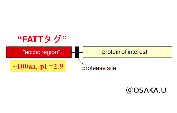 鐚anagawa T (Osaka University) et al developed FATT tag that greatly simplifies purification and refolding procedure at very low cost.
| Technology Developments: Protein Production PPD1: "Development of novel affinity tag system for the high-quality production of extracellular and membrane proteins" (PI) Junichi Takagi | |
| Press Release (in Japanese) from Osaka University | |
| Protein Sci 2013 Mar 23. A multipurpose fusion tag derived from an unstructured and hyperacidic region of the amyloid precursor protein. Sangawa T, Tabata S, Suzuki K, Saheki Y, Tanaka K, Takagi J. |
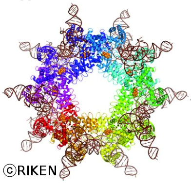 −Itoh Y. et al in RIKEN elucidate the structure and functions of the decameric SelA complex that binds to 10 tRNASec molecules. The molecular weight of the complex is as large as 810 thousand.
| Technology Developments: Protein Production PPC1 “Development of Advanced Production Technologies for Target Proteins” (PI) Shigeyuki Yokoyama | |
| Press Release (in Japanese) from RIKEN | |
| Science 2013 Apr 5;340(6128):75-8. The decameric SelA/tRNASec ring structure reveals the mechanism of bacterial selenocysteine formation. Itoh Y, Brocker MJ, Sekine S, Hammond G, Suetsugu S, Soll D, Yokoyama S. PDB ID: 3W1H,3W1I,3W1J,3W1K |
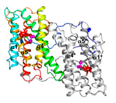 −Inaddition to haem copper oxidases, all higher plants, some algae,yeasts, molds, metazoans, and pathogenic microorganisms such asTrypanosomabrucei contain an additional terminaloxidase, the cyanide-insensitive alternativeoxidase (AOX). AOX is a diiron carboxylate protein that catalyzesthe four-electron reduction of dioxygen to water by ubiquinol.In T. brucei, a parasite that causes human Africansleeping sickness, AOX plays a critical role in the survival ofthe parasite in its bloodstream form. Because AOX is absent frommammals, this protein represents a unique and promising therapeutictarget. Despite its bioenergetic and medical importance, however,structural features of any AOX are yet to be elucidated. KiyoshiKita, Shigeharu Hadada and their colleagues reported crystalstructures of the trypanosomal alternative oxidase in the absence andpresence of ascofuranonederivatives. All structures reveal that the oxidase is a homodimerwith the nonhaem diiron carboxylate active site buried within afour-helix bundle. Unusually, the active site is ligated solely byfour glutamate residues in its oxidized inhibitor-free state;however, inhibitor binding induces the ligation of a histidineresidue. A detailed knowledge of the active site of the enzyme in thepresence of the inhibitors will lead to a greater rational design offurther potent and safer antitrypanosomal drugs.
| Medicine / Pharmacology A5: Development of anti-trypanosome drugs targeting nucleotides biosynthesis and red-ox regulatory pathways (Principal Investigator: Kiyoshi Kita) | |
| Press Release (in Japanese) from U. Tokyo -Kyoto Inst. Technol. | |
| PNAS (2013) March 4 Structure of the trypanosome cyanide-insensitive alternative oxidase Tomoo Shiba, Yasutoshi Kido, Kimitoshi Sakamoto, Daniel Ken, Inaoka, Chiaki Tsuge, Ryoko Tatsumi, Gen Takahashi, Emmanuel Oluwadare Balogun, Takeshi Nara, Takashi Aoki, Teruki Honma, Akiko Tanaka, Masayuki Inoue, Shigeru Matsuoka, Hiroyuki Saimoto, Anthony L. Moore, Shigeharu Harada, and Kiyoshi Kita PDB ID: 3VV9, 3VVA and 3W54 |
 −In various cellular membrane systems, vacuolar ATPases (V-ATPases) function as proton pumps, which are involved in many processes such as bone resorption and cancer metastasis, and these membrane proteins represent attractive drug targets for osteoporosis and cancer. The hydrophilic V1 portion is known as a rotary motor, in which a central axis DF complex rotates inside a hexagonally arranged catalytic A3B3 complex using ATP hydrolysis energy, but the molecular mechanism is not well defined owing to a lack of high-resolution structural information. Takeshi Murata and his colleagues previously reported on the in vitro expression, purification and reconstitution of Enterococcus hirae V1-ATPase from the A3B3 and DF complexes. Now they reported the asymmetric structures of the nucleotide-free and nucleotide-bound A3B3 complex that demonstrate conformational changes induced by nucleotide binding, suggesting a binding order in the right-handed rotational orientation in a cooperative manner. The crystal structures of the nucleotide free and nucleotide-bound V1-ATPase are also reported. The more tightly packed nucleotide-binding site seems to be induced by DF binding, and ATP hydrolysis seems to be stimulated by the approach of a conserved arginine residue. These asymmetric structures represent the first high-resolution view of the rotational mechanism of V1-ATPase.
| Fundamental Biology B4: Towards structure-based design of novel inhibitors for V -ATPase (Principal Investigator: So Iwata) | |
| Press Release (in Japanese) from Chiba U. - JST - RIKEN -Kyoto U. | |
| Nature (2013) January 13 Rotation mechanism of Enterococcus hirae V1-ATPase based on asymmetric crystal structures Satoshi Arai, Shinya Saijo, Kano Suzuki, Kenji Mizutani, Yoshimi Kakinuma, Yoshiko Ishizuka-Katsura, Noboru Ohsawa, Takaho Terada, Mikako Shirouzu, Shigeyuki Yokoyama, So Iwata, Ichiro Yamato & Takeshi Murata PDB ID: 3VR2, 3VR3, 3VR4, 3VR5, and 3VR6 |
 - PIWI-interacting RNAs(piRNAs) silence transposons to maintain genome integrity in animal germ lines. piRNAs are classified as primary and secondary piRNAs, depending on their biogenesis machinery. Primary piRNAs are processed from long non-coding RNA precursors transcribed from piRNA clusters in the genome through the primary processing pathway. Although the existence of a ribonuclease participating in this pathway has been predicted, its molecular identity remained unknown. Osamu Nureki, Haruhiko Siomi and their colleagues showed that Zucchini (Zuc), a mitochondrial phospholipase D superfamily member, is an endoribonuclease essential for primary piRNA biogenesis. The crystal structure of Drosophila melanogaster Zuc (DmZuc) revealed that DmZuc has a positively charged, narrow catalytic groove at the dimer interface, which could accommodate a single-stranded, but not a double-stranded, RNA. DmZuc showed endoribonuclease activity for single-stranded RNAs in vitro.The RNA cleavage products bear a 5′-monophosphate group, a hallmark of mature piRNAs. Mutational analyses revealed that the conserved active-site residues of DmZuc are critical for the ribonuclease activity, piRNA maturation and transposon silencing. They proposed a model for piRNA biogenesis in animal germ lines, in which the Zuc endoribonuclease has a key role in primary piRNA maturation.
| Fundamental Biology B5: Elucidation of the mechanism of high-order cellular functions achieved by non-coding RNAs (Principal Investigator: Osamu Nureki) | |
| Press Release (in Japanese) from JST - U. Tokyo | |
| Nature (2012) October 1 Structure and function of Zucchini endoribonuclease in piRNA biogenesis Hiroshi Nishimasu, Hirotsugu Ishizu, Kuniaki Saito, Satoshi Fukuhara, Miharu K. Kamatani, Luc Bonnefond, Naoki Matsumoto, Tomohiro Nishizawa, Keita Nakanaga, Junken Aoki, Ryuichiro Ishitani, Haruhiko Siomi, Mikiko C. Siomi & Osamu Nureki PDB ID: 4GEL, 4GEM, and 4GEN |
 -Enpp1 is a membrane-bound glycoprotein that regulates bone mineralization by hydrolyzing extracellular nucleotide triphosphates to produce pyrophosphate. Enpp1 dysfunction causes human diseases characterized by ectopic calcification. Enpp1 also inhibits insulin signaling, and an Enpp1 polymorphism is associated with insulin resistance. However, the precise mechanism by which Enpp1 functions in these cellular processes remains elusive. Osamu Nureki, Junken Aoki, Junichi Takagi and their colleagues reported the crystal structures of the extracellular region of mouse Enpp1 in complex with four different nucleotide monophosphates, at resolutions of 2.7–3.2 A. The nucleotides are accommodated in a pocket formed by an insertion loop in the catalytic domain, explaining the preference of Enpp1 for an ATP substrate. Structural mapping of disease-associated mutations indicated the functional importance of the interdomain interactions. A structural comparison of Enpp1 with Enpp2, a lysophospholipase D, revealed marked differences in the domain arrangements and active-site architectures. Notably, the Enpp1 mutant lacking the insertion loop lost the nucleotide-hydrolyzing activity but instead gained the lysophospholipid-hydrolyzing activity of Enpp2. Their findings provide structural insights into how the Enpp family proteins evolved to exert their diverse cellular functions.
| Medicine/Pharmacology B3: Structure and drug development of NPP family members involved in cancer and various diseases (Principal Investigator: Junken Aoki) | |
| Press Release (in Japanese) from U. Tokyo | |
| PNAS (2012) October 1 Crystal structure of Enpp1, an extracellular glycoprotein involved in bone mineralization and insulin signaling Kazuki Kato, Hiroshi Nishimasu, Shinichi Okudaira, Emiko Mihara, Ryuichiro Ishitani, Junichi Takagi, Junken Aoki, and Osamu Nureki PDB ID: 4GTW (AMP complex), 4GTX (TMP complex), 4GTY (GMP complex), and 4GTZ (CMP complex) |
 −Arabidopsis thalianaDEHYDRATION-RESPONSIVE ELEMENT BINDING PROTEIN2A (DREB2A) functions as a
| Food / Environment A3: Structural and functional analyses of transcriptional regulatory proteins useful for breading of drought and heat stress tolerant crops (Principal Investigator: Masaru Tanokura) | |
| Press Release (in Japanese) from U. Tokyo | |
| Plant Cell. (2012) August Arabidopsis GROWTH-REGULATING FACTOR7 Functions as a Transcriptional Repressor of Abscisic Acid-and Osmoti Stress-Responsive Genes, Including DREB2A June-Sik Kim, Junya Mizoi, Satoshi Kidokoro, Kyonoshin Maruyama, Jun Nakajima, Kazuo Nakashima, Nobutaka Mitsuda, Yuko Takiguchi, Masaru Ohme-Takagi, Youichi Kondou, Takeshi Yoshizumi, Minami Matsui, Kazuo Shinozaki, and Kazuko Yamaguchi-Shinozaki |
 - Keap1-Nrf2 system is essential for cytoprotection against oxidative and electrophilic insults. Under unstressed conditions, Keap1 serves as an adaptor for ubiquitin E3 ligase and promotes proteasomal degradation of Nrf2,but Nrf2 is stabilized when Keap1 is inactivated under oxidative stress conditions. Autophagy-deficient mice show aberrant accumulation of p62, a multifunctional scaffold protein, and develop severe liver damage. The p62 accumulation disrupts the Keap1-Nrf2 association and provokes Nrf2 stabilization and accumulation. However, individual contributions of p62 and Nrf2 to the autophagy-deficiency–driven liver pathogenesis have not been clarified. Masayuki Yamamoto and his colleagues revealed that the cellular Keap1 protein levels are increased in Atg7-deficient mouse livers and also in p62-deficient mouse livers, indicating that Keap1 is degraded through the autophagy pathway in a p62-dependent manner. This finding is in clear contrast to the Nrf2 degradation through the proteasome pathway. The Keap1 degradation is accelerated in the presence of certain electrophiles, such as tert-butylhydroquinone (tBHQ). Therefore, the accumulation of Nrf2 and subsequent activation of Nrf2-tartget enzymes is the dominant cause of the liver damage in the autophagy-deficient mice.The autophagy pathway maintains the integrity of the Keap1-Nrf2 system for the normal liver function by governing the Keap1 turnover.
| Fundamental Biology B1: Structural Basis of Sensor System for Cytoprotective Gene Expression Responding to Carcinogens and Oxidative Stress (Principal Investigator: Masayuki Yamamoto) | |
| Press Release (in Japanese) from Tohoku U. | |
| Proc Natl Acad Sci USA. (2012) August 7 Keap1 degradation by autophagy for the maintenance of redox homeostasis Keiko Taguchi, Nanako Fujikawa, Masaaki Komatsu, Tetsuro Ishii, Michiaki Unno, Takaaki Akaike, Hozumi Motohashi, and Masayuki Yamamoto |
 -Cancer cells consume large quantities of nutrients and maintain high levels of anabolism. Recent studies revealed that various oncogenic pathways are involved in modulation of metabolism. Nrf2, a key regulator for the maintenance of redox homeostasis, has been shown to contribute to malignant phenotypes of cancers including aggressive proliferation. However, the mechanisms with which Nrf2 accelerates proliferation are not fully understood. Masayuki Yamamoto, Hozumi Motohashi and their colleagues showed that Nrf2 redirects glucose and glutamine into anabolic pathways, especially under the sustained activation of PI3K-Akt signaling. The active PI3K-Akt pathway augments the nuclear accumulation of Nrf2 and enables Nrf2 to promote metabolic activities that support cell proliferation in addition to enhancing cytoprotection. The functional expansion of Nrf2 reinforces the metabolic reprogramming triggered by proliferative signals
| Fundamental Biology B1: Structural Basis of Sensor System for Cytoprotective Gene Expression Responding to Carcinogens and Oxidative Stress (Principal Investigator: Masayuki Yamamoto) | |
| Press Release (in Japanese) from Tohoku U. - U. Tokyo - National Cancer Center Research Institute | |
| Cancer Cell. (2012) 22, 66-79, July 10 Nrf2 Redirects Glucose and Glutamine into Anabolic Pathways in Metabolic Reprogramming. Yoichiro Mitsuishi, Keiko Taguchi, Yukie Kawatani, Tatsuhiro Shibata, Toshihiro Nukiwa, Hiroyuki Aburatani, Masayuki Yamamoto, Hozumi Motohashi |
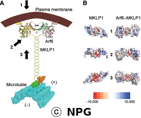 −A small GTPase, Arf6, is involved in cytokinesis by localizing to the Flemming body (the midbody). However, it remains unknown how Arf6 contributes to cytokinesis. Kazuhisa Nakayama, Soichi Wakatsuki and their colleagues demonstrated that Arf6 directly interacts with mitotic kinesin-like protein 1 (MKLP1), a Flemming body-localizing protein essential for cytokinesis. The crystal structure of the Arf6–MKLP1 complex revealed that MKLP1 forms a homodimer flanked by two Arf6 molecules, forming a 2:2 heterotetramer containing an extended β-sheet composed of 22 β-strands that spans the entire heterotetramer, suitable for interaction with a concave membrane surface at the cleavage furrow. They showed that, during cytokinesis, Arf6 is first accumulated around the cleavage furrow and, prior to abscission, recruited onto the Flemming body via interaction with MKLP1. They also showed by structure-based mutagenesis and siRNA-mediated knockdowns that the complex formation is required for completion of cytokinesis. A model based on these results suggests that the Arf6–MKLP1 complex plays a crucial role in cytokinesis by connecting the microtubule bundle and membranes at the cleavage plane.
| Fundamental Biology A6: Structure-function analysis of protein complexes that regulate vesicular traffic (Principal Investigator: Soichi Wakatsuki) | |
| Press Release(in Japanese) from Kyoto U. | |
| EMBO J. (2012) 31, 2590-2603 Structural basis for Arf6-MKLP1 complex formation on the Flemming body responsible for cytokinesis. Makyio H, Ohgi M, Takei T, Takahashi S, Takatsu H, Katoh Y, Hanai A, Ueda T, Kanaho Y, Xie Y, Shin HW, Kamikubo H, Kataoka M, Kato R, Wakatsuki S, Nakayama K. PDB ID: 3VHX |
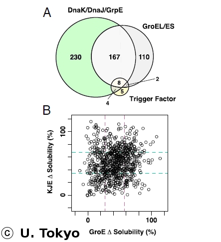 −Protein folding is
often hampered by protein aggregation, which can be prevented by a variety of chaperones in the cell. A dataset that evaluates which chaperones are effective for aggregation-prone proteins would provide an invaluable resource not only for understanding the roles of chaperones, but also for broader applications in protein science and engineering. Takuya Ueda and his colleagues comprehensively evaluated the effects of the major Escherichia colichaperones, trigger factor, DnaK/DnaJ/GrpE, and GroEL/GroES, on 800 aggregation prone cytosolic E. coli proteins, using a reconstituted chaperone-free translation system. Statistical analyses revealed the robustness and the intriguing properties of chaperones. The DnaK and GroEL systems drastically increased the solubilities of hundreds of proteins with weak biases, whereas trigger factor had only a marginal effect on solubility. The combined addition of the chaperones was effective for a subsetf proteins that were not rescued by any single chaperone system, supporting the synergistic effect of these chaperones. The resource, which is accessible via a public database, eSol, can be used to investigate the properties of proteins of interest in terms of their solubilities and chaperone effects.
−Protein folding is
often hampered by protein aggregation, which can be prevented by a variety of chaperones in the cell. A dataset that evaluates which chaperones are effective for aggregation-prone proteins would provide an invaluable resource not only for understanding the roles of chaperones, but also for broader applications in protein science and engineering. Takuya Ueda and his colleagues comprehensively evaluated the effects of the major Escherichia colichaperones, trigger factor, DnaK/DnaJ/GrpE, and GroEL/GroES, on 800 aggregation prone cytosolic E. coli proteins, using a reconstituted chaperone-free translation system. Statistical analyses revealed the robustness and the intriguing properties of chaperones. The DnaK and GroEL systems drastically increased the solubilities of hundreds of proteins with weak biases, whereas trigger factor had only a marginal effect on solubility. The combined addition of the chaperones was effective for a subsetf proteins that were not rescued by any single chaperone system, supporting the synergistic effect of these chaperones. The resource, which is accessible via a public database, eSol, can be used to investigate the properties of proteins of interest in terms of their solubilities and chaperone effects.
| Protein Production C1: Development of Advanced Production Technologies for Target Proteins (Principal Investigator: Shigeyuki Yokoyama) | |
| Press Release(in Japanese) from U. Tokyo | |
| Proc Natl Acad Sci USA, 2012 May 21 Global analysis of chaperone effects using a reconstituted cell-free translation system. Niwa T, Kanamori T, Ueda T, Taguchi H. |
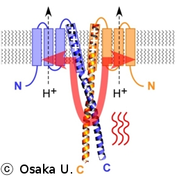 −Hv1/VSOP is a dimeric voltage-gated H+ channel in which the gating of one subunit is reportedly coupled to that of the other subunit within the dimer. The molecular basis for dimer formation and intersubunit coupling, however, remains unknown. Yasushi Okamura and his colleagues showed that the carboxy terminus ends downstream of the S4 voltage-sensor helix twist in a dimer coiled-coil architecture, which mediates cooperative gating. They also showed that the temperature-dependent activation of H+ current through Hv1/VSOP is regulated by thermostability of the coiled-coil domain, and that this regulation is altered by mutation of the linker between S4 and the coiled-coil. Cooperative gating within the dimer is also dependent on the linker structure, which circular dichroism spectrum analysis suggests is α-helical. Their results indicate that the cytoplasmic coiled-coil strands form continuous α-helices with S4 and mediate cooperative gating to adjust the range of temperatures over which Hv1/VSOP operates.
| Fundamental Biology A5: Structure and function of voltage-sensor domain proteins (Principal Investigator: Yasushi Okamura) | |
| Press Release(in Japanese) from Osaka U. | |
| Nature Communications, 2012 May 9 The cytoplasmic coiled-coil mediates cooperative gating temperature sensitivity in the voltage-gated H+ channel Hv1 Yuichiro Fujiwara, Tatsuki Kurokawa, Kohei Takeshita, Megumi Kobayashi, Yoshifumi Okochi, Atsushi Nakagawa, Yasushi Okamura PDB ID: 3VMX |
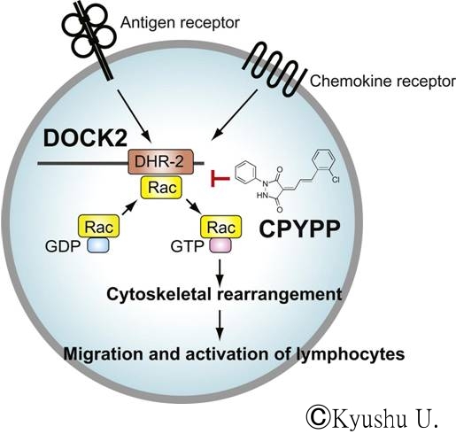 −Tissue infiltration of activated lymphocytes is a hallmark of transplant rejection and organ-specific autoimmune diseases. Migration and activation of lymphocytes depend on DOCK2, an atypical Rac activator predominantly expressed in hematopoietic cells. Although DOCK2 does not contain Dbl homology domain typically found in guanine nucleotide exchange factors, DOCK2 mediates the GTP-GDP exchange reaction for Rac through its DHR-2 domain. Yoshinori Fukui and his colleagues identified 4-[3′-(2″-chlorophenyl)-2′-propen-1′-ylidene]-1-phenyl-3,5-pyrazolidinedione (CPYPP) as a small-molecule inhibitor of DOCK2. CPYPP bound to DOCK2 DHR-2 domain in a reversible manner and inhibited its catalytic activity in vitro. When lymphocytes were treated with CPYPP, both chemokine receptor- and antigen receptor-mediated Rac activation were blocked, resulting in marked reduction of chemotactic response and T cell activation. These results provide a rational
of and a chemical scaffold for development of the DOCK2-targeting immunosuppressant.
−Tissue infiltration of activated lymphocytes is a hallmark of transplant rejection and organ-specific autoimmune diseases. Migration and activation of lymphocytes depend on DOCK2, an atypical Rac activator predominantly expressed in hematopoietic cells. Although DOCK2 does not contain Dbl homology domain typically found in guanine nucleotide exchange factors, DOCK2 mediates the GTP-GDP exchange reaction for Rac through its DHR-2 domain. Yoshinori Fukui and his colleagues identified 4-[3′-(2″-chlorophenyl)-2′-propen-1′-ylidene]-1-phenyl-3,5-pyrazolidinedione (CPYPP) as a small-molecule inhibitor of DOCK2. CPYPP bound to DOCK2 DHR-2 domain in a reversible manner and inhibited its catalytic activity in vitro. When lymphocytes were treated with CPYPP, both chemokine receptor- and antigen receptor-mediated Rac activation were blocked, resulting in marked reduction of chemotactic response and T cell activation. These results provide a rational
of and a chemical scaffold for development of the DOCK2-targeting immunosuppressant.
| Medicine and PharmacologyA2: Drug discovery-oriented analysis for structure and function of DOCK2 signaling molecules (Principal Investigator: Yoshinori Fukui) | |
| Press Release(in Japanese) from Kyushu U.- JST | |
| Chemistry and Biology 2012 April 20 Blockade of Inflammatory Responses by a Small-Molecule Inhibitor of the Rac Activator DOCK2 Akihiko Nishikimi, Takehito Uruno, Xuefeng Duan, Qinhong Cao, Yuji Okamura, Takashi Saitoh, Nae Saito, Shunsuke Sakaoka, Yao Du, Atsushi Suenaga, Mutsuko Kukimoto-Niino, Kei Miyano, Kazuhito Gotoh, Takayoshi Okabe, Fumiyuki Sanematsu, Yoshihiko Tanaka, Hideki Sumimoto, Teruki Honma, Shigeyuki Yokoyama, Tetsuo Nagano, Daisuke Kohda, Motomu Kanai, Yoshinori Fukui |
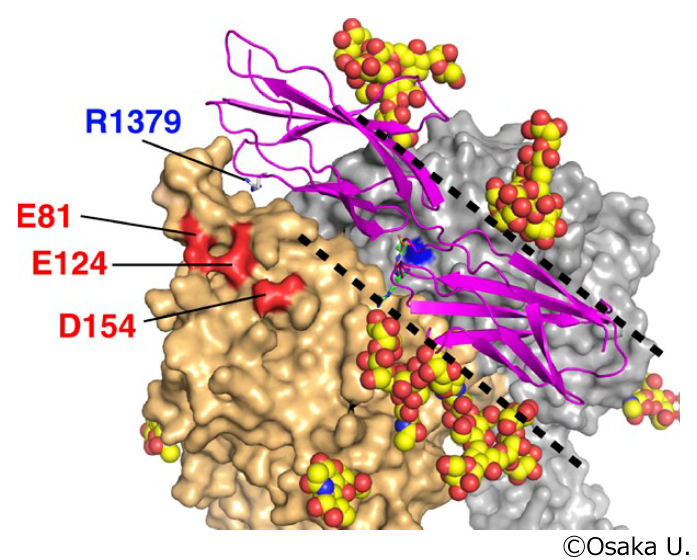 −Integrin α5 β1 is a major cellular receptor for the extracellular matrix protein fibronectin and plays a fundamental role during mammalian development. Junichi Takagi and his colleagues determined a crystal structure of the α5β1 integrin headpiece fragment bound by an allosteric inhibitory antibody at a 2.9-A resolution both in the absence and presence of a ligand peptide containing the Arg-Gly-Asp (RGD) sequence. The antibody-bound β1 chain accommodated the RGD ligand with very limited structural changes, which may represent the initial step of cell adhesion mediated by nonactivated integrins. Furthermore, a molecular dynamics simulation pointed to an important role for Ca2+ in the conformational coupling between the ligand-binding site and the rest of the molecule. The RGD-binding pocket is situated at the center of a trenchlike exposed surface on the top face of α5β1 devoid of glycosylation sites. The structure also enabled the precise prediction of the acceptor residue for the auxiliary synergy site of fibronectin on the α5 subunit, which was experimentally confirmed by mutagenesis and kinetic binding assays.
| Protein Production D1: Development of novel affinity tag system for the high-quality production of extracellular and membrane proteins (Principal Investigator: Junichi Takagi) | |
| Press Release(in Japanese) from Osaka U. | |
| J. Cell Biol, 2012 March 26 Crystal structure of α5β1 integrin ectodomain: Atomic details of the fibronectin receptor Masamichi Nagae, Suyong Re, Emiko Mihara, Terukazu Nogi, Yuji Sugita, and Junichi Takagi PDB ID: 3VI3, 3VI4 |
 −Free fatty acids provide an important energy source as nutrients, and act as signaling molecules in various cellular processes. Several G-protein-coupled receptors have been identified as free-fatty-acid receptors important in physiology as well as in several diseases. GPR120 (also known as O3FAR1) functions as a receptor for unsaturated long-chain free fatty acids and has a critical role in various physiological homeostasis mechanisms such as adipogenesis, regulation of appetite and food preference. Gozoh Tsujimoto and his colleagues at Kyoto U. and in Europe showed that GPR120-deficient mice fed a high-fat diet develop obesity, glucose intolerance and fatty liver with decreased adipocyte differentiation and
lipogenesis and enhanced hepatic lipogenesis. Insulin resistance in such mice is associated with reduced insulin signaling and enhanced inflammation in adipose tissue. In human, we show that GPR120 expression in adipose tissue is significantly higher in obese individuals than in lean controls. GPR120 exon sequencing in obese subjects reveals a deleterious non-synonymous mutation (p.R270H) that inhibits GPR120 signaling activity. Furthermore, the p.R270H variant increases the risk of obesity in European populations. Overall, their study demonstrated that the lipid sensor GPR120 has a key role in sensing dietary fat and, therefore, in the control of energy balance in both humans and rodents.
−Free fatty acids provide an important energy source as nutrients, and act as signaling molecules in various cellular processes. Several G-protein-coupled receptors have been identified as free-fatty-acid receptors important in physiology as well as in several diseases. GPR120 (also known as O3FAR1) functions as a receptor for unsaturated long-chain free fatty acids and has a critical role in various physiological homeostasis mechanisms such as adipogenesis, regulation of appetite and food preference. Gozoh Tsujimoto and his colleagues at Kyoto U. and in Europe showed that GPR120-deficient mice fed a high-fat diet develop obesity, glucose intolerance and fatty liver with decreased adipocyte differentiation and
lipogenesis and enhanced hepatic lipogenesis. Insulin resistance in such mice is associated with reduced insulin signaling and enhanced inflammation in adipose tissue. In human, we show that GPR120 expression in adipose tissue is significantly higher in obese individuals than in lean controls. GPR120 exon sequencing in obese subjects reveals a deleterious non-synonymous mutation (p.R270H) that inhibits GPR120 signaling activity. Furthermore, the p.R270H variant increases the risk of obesity in European populations. Overall, their study demonstrated that the lipid sensor GPR120 has a key role in sensing dietary fat and, therefore, in the control of energy balance in both humans and rodents.
| Chemical Regulation C1: Establishment of Chemical Library and Development of Protein Regulation Technology (Principal Investigator: Tetsuo Nagano) | |
| Faulty fat sensor implicated in obesity and liver disease from Imperial College London | |
| Nature, 2012 February 19 Dysfunction of lipid sensor GPR120 leads to obesity in both mouse and human Atsuhiko Ichimura, Akira Hirasawa, Odile Poulain-Godefroy, Amelie Bonnefond, Takafumi Hara, Loic Yengo, Ikuo Kimura, Audrey Leloire, Ning Liu, Keiko Iida, Helene Choquet, Philippe Besnard, Cecile Lecoeur, Sidonie Vivequin, Kumiko Ayukawa, Masato Takeuchi, Kentaro Ozawa, Maithe Tauber, Claudio Maffeis, Anita Morandi,Raffaella Buzzetti, Paul Elliott, Anneli Pouta, Marjo-Riitta Jarvelin, Antje Korner, Wieland Kiess, Marie Pigeyre, Roberto Caiazzo, Wim Van Hul, Luc Van Gaal, Fritz Horber,Beverley Balkau, Claire Levy-Marchal, Konstantinos Rouskas, Anastasia Kouvatsi, Johannes Hebebrand, Anke Hinney, Andre Scherag, Francois Pattou, David Meyre,Taka-aki Koshimizu, Isabelle Wolowczuk, Gozoh Tsujimoto, Philippe Froguel |
 −DOCK2(dedicator of cytokinesis 2) controls lymphocyte migration through ras-related C3 botulinum toxin substrate (Rac) activation. DOCK2-engulfment and cell motility protein 1 (DOCK2・ELMO1) complex formation is required for DOCK2-mediated Rac signaling. Shigeki Yokoyama, Yoshinori Fukui, and their colleagues identified the N-terminal 177-residue fragment and the C-terminal 196-residue fragment of human DOCK2 and ELMO1, respectively, as the mutual binding regions, and solved the crystal structure of their complex at 2.1-A resolution. Overall, the entire regions of both DOCK2 and ELMO1 assemble to create a rigid structure, which is required for the DOCK2・ELMO1 binding, as revealed by mutagenesis. We demonstrated that the ELMO-interacting region and the DOCK-homology region 2 guanine nucleotide exchange factor domain of DOCK2 associate with each other for the autoinhibition, and that the assembly with ELMO1 weakens the interaction, relieving DOCK2 from the autoinhibition. Therefore, the DOCK2?ELMO1 complex structure reveals the structural basis by which DOCK2 and ELMO1 mutually relieve their autoinhibition for the activation of Rac1 for lymphocyte chemotaxis.
|
Protein Production C1: Development of Advanced Production Technologies for Target Proteins (Principal Investigator: Shigeyuki Yokoyama) Medicine/Pharmacology A2: Drug discovery-oriented analysis for structure and function of DOCK2 signaling molecules (Principal Investigator: Yoshinori Fukui) TP Atlas |
|
| Press Release(in Japanese) from RIKEN - Kyushu U. | |
| PNAS, 2012 February 13 Structural basis for mutual relief of the Rac guanine nucleotide exchange factor DOCK2 and its partner ELMO1 from their autoinhibited forms Kyoko Hanawa-Suetsugu, Mutsuko Kukimoto-Niino, Chiemi Mishima-Tsumagari, Ryogo Akasaka, Noboru Ohsawa, Shun-ichi Sekine, Takuhiro Ito, Naoya Tochio, Seizo Koshiba, Takanori Kigawa, Takaho Terada, Mikako Shirouzu, Akihiko Nishikimi, Takehito Uruno, Tomoya Katakai, Tatsuo Kinashi, Daisuke Kohda, Yoshinori Fukui and Shigeyuki Yokoyama PDB ID: 2RQR, 3A98, 3B13 |
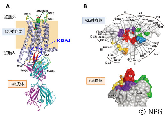 −G-protein-coupled receptors are the largest class of cell-surface receptors, and these membrane proteins exist in equilibrium between inactive and active states. Conformational changes induced by extracellular ligands binding to G-protein-coupled receptors result in a cellular response through the activation of G proteins. The A2A adenosine receptor (A2AAR) is responsible for regulating blood flow to the cardiac muscle and is important in the regulation of glutamate and dopamine release in the brain. So Iwata and his colleagues reported the raising of a mouse monoclonal antibody against human A2AAR that prevents agonist but not antagonist binding to the extracellular ligand-binding pocket, and describe the structure of A2AAR in complex with the antibody Fab fragment (Fab2838). The structure reveals that Fab2838 recognizes the intracellular surface of A2AAR and that its complementarity-determining region, CDR-H3, penetrates into the receptor. CDR-H3 is located in a similar position to the G-protein carboxy-terminal fragment in the active opsin structure and to CDR-3 of the nanobody in the active β2-adrenergic receptor structure, but locks A2AAR in an inactive conformation. Their results suggest a new strategy to modulate the activity of G-protein-coupled receptors.
|
Fundamental Biology B4: Membrane transporters: structure and function of important drug targets (Principal Investigator: So Iwata) TP Atlas Protein Production D3: Antibody production for Membrane Protein Crystallization (Principal Investigator: So Iwata) |
|
| Press Release(in Japanese) from JST - Kyoto U. - U. Tokyo - Chiba U. | |
| Nature, 2012 January 29 G-protein-coupled receptor inactivation by an allosteric inverse-agonist antibody Tomoya Hino, Takatoshi Arakawa, Hiroko Iwanari, Takami Yurugi-Kobayashi, Chiyo Ikeda-Suno, Yoshiko Nakada-Nakura, Osamu Kusano-Arai, Simone Weyand, Tatsuro Shimamura, Norimichi Nomura, Alexander D. Cameron, Takuya Kobayashi, Takao Hamakubo, So Iwata & Takeshi Murata PDB ID: 3VG9, 3VGA |
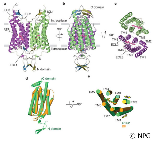 −Channelrhodopsins (ChRs) are light-gated cation channels derived from algae that have shown experimental utility in optogenetics; for example, neurons expressing ChRs can be optically controlled with high temporal precision within systems as complex as freely moving mammals. Although ChRs have been broadly applied to neuroscience research, little is known about the molecular mechanisms by which these unusual and powerful proteins operate. Osamu Nureki and his colleagues presented the crystal structure of a ChR (a C1C2 chimaera between ChR1 and ChR2 from Chlamydomonas reinhardtii) at 2.3A? resolution using the beamline BL32XU of Spring-8 developed in the TPRP. The structure reveals the essential molecular architecture of ChRs, including the retinal-binding pocket and cation conduction pathway. The integration of structural and electrophysiological analyses provides insight into the molecular basis for the remarkable function of ChRs, and paves the way for the precise and principled design of ChR variants with novel properties.
|
Fundamental Biology B5: Elucidation of the mechanism of high-order cellular functions achieved by non-coding RNAs (Principal Investigator: Osamu Nureki) TP Atlas |
|
|
All cations lead to this pathway - The structure of channelrhodopsin reveals its cation-conducting pathway and activation mechanism U. Tokyo | |
| Nature, 2012 January 23 Crystal structure of the channelrhodopsin light-gated cation channel. Kato HE, Zhang F, Yizhar O, Ramakrishnan C, Nishizawa T, Hirata K, Ito J, Aita Y, Tsukazaki T, Hayashi S, Hegemann P, Maturana AD, Ishitani R, Deisseroth K, Nureki O. PDB ID: 3UG9 |
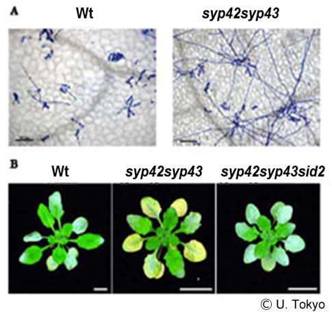 −In all eukaryotic cells, a post-membrane-trafficking system connects the Golgi organelles, such as the trans-Golgi network (TGN), endosomes, vacuoles, and the plasma membrane. This complex network plays critical roles in several higher-order functions in multicellular organisms. The TGN, one of the important organelles for protein transport in the post-Golgi network, functions as a sorting station, where cargo proteins are directed to the appropriate post-Golgi compartments. Unlike its roles in animal and yeast cells, the TGN has also been reported to function like early endosomal compartments in plant cells. However, the physiological roles of the TGN functions in plants are not understood. Akihiko Nakano and his colleagues reported a study of the SYP4 group (SYP41, SYP42, and SYP43), which represents the plant orthologs of the Tlg2/syntaxin16 Qa-SNARE that localizes on the TGN in yeast and animal cells. The SYP4 group regulates the secretory and vacuolar transport pathways in the post-Golgi network and maintains the morphology of the Golgi apparatus and TGN. Consistent with a secretory role, SYP4 proteins are required for extracellular resistance responses to a fungal pathogen. They also revealed a plant cell-specific higher-order role of the SYP4 group in the protection of chloroplasts from salicylic acid-dependent biotic stress.
|
Fundamental Biology A6: Structure-function analysis of protein complexes that regulate vesicular traffic (Principal Investigator: Soichi Wakatsuki) TP Atlas |
|
|
All cations lead to this pathway - The structure of channelrhodopsin reveals its cation-conducting pathway and activation mechanism U. Tokyo | |
| PNAS, January 17, 2012 Qa-SNAREs localized to the trans-Golgi network regulate multiple transport pathways and extracellular disease resistance in plants Tomohiro Uemura, Hyeran Kim, Chieko Saito, Kazuo Ebine, Takashi Ueda, Paul Schulze-Lefert, and Akihiko Nakano |
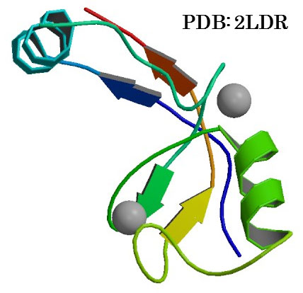 −Cbl-b is a RING-type E3 ubiquitin ligase that functions as a negative regulator of T-cell activation and growth factor receptor and nonreceptor-type tyrosine kinase signaling. Cbl-b dysfunction is related to autoimmune diseases and cancers in humans. However, the molecular mechanism regulating its E3 activity is largely unknown. Using NMR and small-angle X-ray scattering analyses, Fuyuhiko Inagaki and his colleagues revealed that the unphosphorylated N-terminal region of Cbl-b forms a compact structure by an intramolecular interaction, which masks the interaction surface of the RING domain with an E2 ubiquitin-conjugating enzyme. Phosphorylation of Y363, located in the helix-linker region between the tyrosine kinase binding and the RING domains, disrupts the interdomain interaction to expose the E2 binding surface of the RING domain. Structural analysis revealed that the phosphorylated helix-RING region forms a compact structure in solution. Moreover, the phosphate group of pY363 is located in the vicinity of the interaction surface with UbcH5B to increase affinity by reducing their electrostatic repulsion. Thus, the phosphorylation of Y363 regulates the E3 activity of Cbl-b by two mechanisms: one is to remove the masking of the RING domain from the tyrosine kinase binding domain and the other is to form a surface to enhance binding affinity to E2.
|
Fundamental Biology A3: Structural basis of Atg proteins essential for autophagy (Principal Investigator: Fuyuhiko Inagaki) TP Atlas |
|
| Press Release(in Japanese) from Hokkaido U. | |
| PNAS, 2011 December 7 Autoinhibition and phosphorylation-induced activation mechanisms of human cancer and autoimmune disease-related E3 protein Cbl-b Yoshihiro Kobashigawa, Akira Tomitaka, Hiroyuki Kumeta, Nobuo N.Noda, Masaya Yamaguchi, and Fuyuhiko Inagaki PDB ID: 2LDR and 3VGO |
 −E1 enzymes activate ubiquitin-like proteins and transfer them to cognate E2 enzymes. Atg7, a non-canonical E1, activates two ubiquitin-like proteins Atg8 and Atg12, and plays a crucial role in autophagy. Huyuhiko Inagaki and his colleagues reported crystal structures of full-length Atg7 and its C-terminal domain bound to Atg8 and MgATP, as well as a solution structure of Atg8 bound to the extreme C-terminal domain (ECTD) of Atg7. The unique N-terminal domain (NTD) of Atg7 is responsible for Atg3 (E2) binding, whereas its C-terminal domain is comprised of a homodimeric adenylation domain (AD) and ECTD. The structural and biochemical data demonstrate that Atg8 is initially recognized by the C-terminal tail of ECTD, then transferred to AD where the Atg8 C-terminus is attacked by the catalytic cysteine to form a thioester bond. Atg8 is then transferred via a trans mechanism to the Atg3 bound to the NTD of the opposite protomer within a dimer.
|
Fundamental Biology A3: Structural basis of Atg proteins essential for autophagy (Principal Investigator: Fuyuhiko Inagaki) TP Atlas |
|
| Press Release(in Japanese) from Hokkaido U. | |
| Mol Cell., 2011 November 4 Structural basis of Atg8 activation by a homodimeric E1, Atg7. Noda NN, Satoo K, Fujioka Y, Kumeta H, Ogura K, Nakatogawa H,Ohsumi Y, Inagaki F. PDB ID: 3VH1, 3VH2, 3VH3, 3VH4 and 2LI5 |
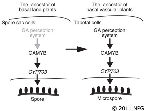 −Gibberellin (GA) controls pollen development in flowering plants via the GAMYB transcription factor. Makoto Matsuoka and his colleagues showed that GAMYB is conserved in Selaginella moellendorffii (lycophyte) and Physcomitrella patens (moss), although the former contains the GA signaling pathway, the latter does not. In the lycophyte, GA treatment promotes the outer wall development on microspores, whereas treatment with GA biosynthesis inhibitors disturbs its development. Contrary, in the moss, GAMYB homologue knockouts also produce abnormal spores that resemble Selaginella microspores treated with GA biosynthesis inhibitors and pollen grains of rice gamyb mutant. Moreover, the knockouts fail to develop male organs, instead ectopically forming female organs. Thus, before the establishment of the GA signaling pathway, basal land plants, including mosses, contained a GAMYB-based system for spore and sexual organ development. Subsequently, during the evolution from mosses to basal vascular plants including lycophytes, GA signaling might have merged to regulate this pre-existing GAMYB-based system.
|
Food and Environment A4: Structural and functional analyses of regulatory proteins in plant growth and stress resistance (Principal Investigator: Makoto Matsuoka) TP Atlas |
|
| Press Release(in Japanese) from Nagoya U.‐NIBB | |
| Nature Communications, 2011 November 22 The Gibberellin perception system evolved to regulate a pre-existing GAMYB-mediated system during land plant evolution Koichiro Aya, Yuji Hiwatashi, Mikiko Kojima, Hitoshi Sakakibara, Miyako Ueguchi-Tanaka, Mitsuyasu Hasebe & Makoto Matsuoka |
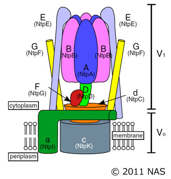 −V-ATPases function as ATP-dependent ion pumps in various membrane systems of living organisms. ATP hydrolysis causes rotation of the central rotor complex, which is composed of the central axis D subunit and a membrane c ring that are connected by F and d subunits. Takeshi Murata and his colleagues determined the crystal structure of the DF complex of the prokaryotic V-ATPase of Enterococcus hirae. The structure of the D subunit comprised a long left-handed coiled coil with a unique short β-hairpin region that is effective in stimulating the ATPase activity of V1-ATPase by twofold. The F subunit is bound to the middle portion of the D subunit. The C-terminal helix of the F subunit, which was believed to function as a regulatory region by extending into the catalytic A3B3 complex, contributes to tight binding to the D subunit by forming a three-helix bundle. Both D and F subunits are necessary to bind the d subunit that links to the c ring. From these findings, they modeled the entire rotor complex (DFdc ring) of V-ATPase.
|
Fundamental Biology B4: Membrane transporters: structure and function of important drug targets (Principal Investigator: So Iwata) TP Atlas |
|
| Press ReleasePress Release (in Japanese) from Chiba U.‐Kyoto U.‐RIKEN | |
| PNAS, 2011 November 23 Crystal structure of the central axis DF complex of the prokaryotic V-ATPase Shinya Saijo, Satoshi Arai, K. M. Mozaffor Hossain, Ichiro Yamato, Kano Suzuki, Yoshimi Kakinuma, Yoshiko Ishizuka-Katsura, Noboru Ohsawa, Takaho Terada, Mikako Shirouzu, Shigeyuki Yokoyama, So Iwata, and Takeshi Murata PDB ID: 3AON |
 −Histones are subjected to a variety of post-translational modifications, such as methylation of lysine and arginine. These modifications regulate the structure and function of chromatin. Tri- and dimethylations of histone H3K27 (H3K27me3/2) represses key developmental genes. The mechanisms by which histone-modifying enzymes selectively regulate the methylation states of H3K27 are poorly understood. Toru Sengoku and Shigeyuki Yokoyama reported the crystal structures of the catalytic fragment of UTX, an H3K27me3/2-specific demethylase, in the free and H3 peptide-bound forms. The catalytic jumonji domain binds H3 residues 25?33, recognizing H3R26, H3A29, and H3P30 in a sequence-specific manner, in addition to H3K27me3 in the catalytic pocket. A novel zinc-binding domain binds residues 17?21 of H3. Mutational analyses showed that H3R17, H3L20, H3R26, H3A29, H3P30, and H3T32 are each important for demethylation. No other methyllysines in the histone tails have the same set of residues at the corresponding positions. Thus, they clarified how UTX discriminates H3K27me3/2 from the other methyllysines with distinct roles, including the near-cognate H3K9me3/2, in histones.
|
Protein Production C1: Development of Advanced Production Technologies for Target Proteins (Principal Investigator: Shigeyuki Yokoyama) |
|
| Press Release(in Japanese) from RIKEN | |
| Genes & Development, 2011, October 16 Structural basis for histone H3 Lys 27 demethylation by UTX/KDM6A Toru Sengoku and Shigeyuki Yokoyama PDB ID: 3AVS, 3AVR |
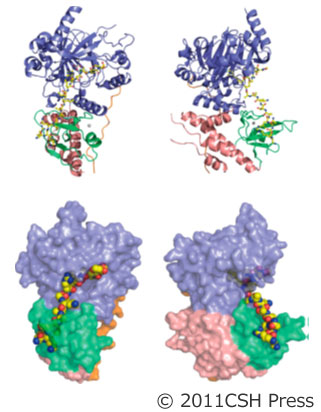
 −Adenomatous polyposis coli (APC) is a tumor suppressor protein commonly mutated in colorectal tumors. APC plays important roles in Wnt signaling and other cellular processes. Shigeyuki Yokoyama at RIKEN, Tetsu Akiyama at U. Tokyo and their colleagues reported the crystal structure of the armadillo repeat (Arm) domain of APC, which facilitates the binding of APC to various proteins. APC-Arm forms a superhelix with a positively charged groove. They also determined the structure of the complex of APC-Arm with the tyrosine-rich (YY) domain of the Src-associated in mitosis, 68 kDa protein (Sam68), which regulates TCF-1 alternative splicing. Sam68-YY forms numerous interactions with the residues on the groove and is thereby fixed in a bent conformation. They assessed the effects of mutations and phosphorylation on complex formation between APC-Arm and Sam68-YY. Structural comparisons revealed different modes of ligand recognition between the Arm domains of APC and other Arm-containing proteins.
| Protein Production C1: Development of Advanced Production Technologies for Target Proteins (Principal Investigator: Shigeyuki Yokoyama) | |
| Press Release (in Japanese) from RIKEN−U. Tokyo | |
| Structure, 2011, October 11 Crystal Structures of the Armadillo Repeat Domain of Adenomatous Polyposis Coli and Its Complex with the Tyrosine-Rich Domain of Sam68. Ella Czarina Morishita, Kazutaka Murayama, Miyuki Kato-Murayama, Yoshiko Ishizuka-Katsura, Yuri Tomabechi, Tomoatsu Hayashi, Takaho Terada, Noriko Handa, Mikako Shirouzu, Tetsu Akiyama, and Shigeyuki Yokoyama PDB ID: 3AU3, 3QHE |
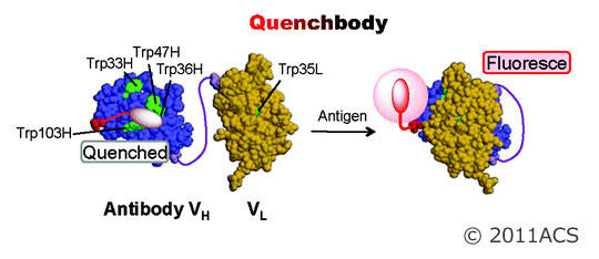 −Hiroshi Ueda, Hiroaki Takagi and their colleagues reported novel reagentless fluorescent biosensor strategy based on the antigen-dependent removal of a quenching effect on a fluorophore attached to antibody domains. Using a cell-free translation-mediated position-specific protein labeling system, they found that an antibody single chain variable region (scFv) that had been fluorolabeled at the N-terminal region showed a significant antigen-dependent fluorescence enhancement. Investigation of the enhancement mechanism by mutagenesis of the carboxytetramethylrhodamine (TAMRA)-labeled anti-osteocalcin scFv showed that antigen-dependency was dependent on semiconserved tryptophan residues near the VH/VL interface. This suggested that the binding of the antigen led to the interruption of a quenching effect caused by the proximity of tryptophan residues to the linker-tagged fluorophore. Using TAMRA-scFv, many targets including peptides, proteins, and haptens including morphine-related drugs could be quantified. Similar or higher sensitivities to those observed in competitive ELISA were obtained, even in human plasma. Because of its versatility, this “quenchbody” is expected to have a range of applications, from in vitro diagnostics, to imaging of various targets in situ.
| Protein Production C1: Development of Advanced Production Technologies for Target Proteins (Principal Investigator: Shigeyuki Yokoyama) | |
| Press Release (in Japanese) from U. Tokyo−JAIST | |
| J. Am. Chem. Soc.,2011, October 6 “Quenchbodies”: Quench-Based Antibody Probes That Show Antigen-Dependent Fluorescence Ryoji Abe, Hiroyuki Ohashi, Issei Iijima, Masaki Ihara, Hiroaki Takagi, Takahiro Hohsaka, and Hiroshi Ueda |
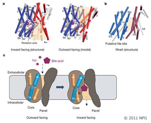 −High cholesterol levels greatly increase the risk of cardiovascular disease. About 50 per cent of cholesterol is eliminated from the body by its conversion into bile acids. However, bile acids released from the bile duct are constantly recycled, being reabsorbed in the intestine by the apical sodium-dependent bile acid transporter (ASBT, also known as SLC10A2). It has been shown in animal models that plasma cholesterol levels are considerably lowered by specific inhibitors of ASBT and ASBT is thus a target for hypercholesterolaemia drugs. So Iwata and his colleagues reported the crystal structure of a bacterial homologue of ASBT from Neisseria meningitides (ASBTNM). ASBTNM contains two inverted structural repeats of five transmembrane helices. A core domain of six helices harbors two sodium ions, and the remaining four helices pack in a row to form a flat, ‘panel’-like domain. The ASBTNM structure was captured with the substrate taurocholate present, bound between the core and panel domains in a large, inward-facing, hydrophobic cavity. Residues near this cavity have been shown to affect the binding of specific inhibitors of human ASBT4. The position of the taurocholate molecule, together with the molecular architecture, suggests the rudiments of a possible transport mechanism.
| Fundamental Biology B4: Membrane transporters: structure and function of important drug targets (Principal Investigator: So Iwata) TP Atlas |
|
| Press Release (in Japanese) from JST−Kyoto U. | |
| Nature2011 October 5 Crystal structure of a bacterial homologue of the bile acid sodium symporter ASBT. Hu NJ, Iwata S, Cameron AD, Drew D. PDB ID: 3ZUY, 3ZUX |
 −Seasonal fluctuations in day length regulate important aspects of plant development such as the flowering transition or, in potato (Solanum tuberosum), the formation of tubers. Day length is sensed by the leaves, which produce a mobile signal transported to the shoot apex or underground stems to induce a flowering transition or, respectively, a tuberization transition. Work in Arabidopsis, tomato and rice (Oryza sativa) identified the mobile FLOWERING LOCUS T (FT) protein as a main component of the long-range ‘florigen’, or flowering hormone, signal. Ko Shimamoto at NAIST and his colleagues have shown show that expression of the Hd3a gene, the FT orthologue in rice, induces strict short-day potato types to tuberize in long days. Tuber induction is graft transmissible and the Hd3a-GFP protein is detected in the stolons of grafted plants, transport of the fusion protein thus correlating with tuber formation. They provide evidence showing that the potato floral and tuberization transitions are controlled by two different FT-like paralogues (StSP3D and StSP6A) that respond to independent environmental cues, and show that an autorelay mechanism involving CONSTANS modulates expression of the tuberization-control StSP6A gene.
| Food / Environment B6 | |
| Food / Environment B6: Structure and function of environmentally-responsive proteins (Principal Investigator: Ko Shimamoto) TP Atlas |
|
| Press Release (in Japanes) from NAIST | |
| Nature, 2011 September 25 Control of flowering and storage organ formation in potato by FLOWERING LOCUS T. Navarro C, Abelenda JA, Cruz-Oro E, Cuellar CA, Tamaki S, Silva J, Shimamoto K, Prat S. |
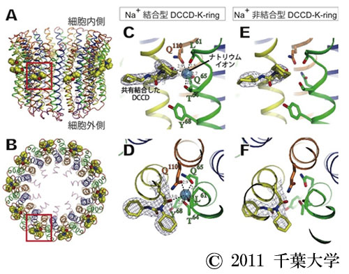 − The prokaryotic V-ATPase of Enterococcus hirae, closely related to the eukaryotic enzymes, provides a unique opportunity to study the ion-translocation mechanism because it transports Na+, which can be detected by radioisotope (22Na+ ) experiments and X-ray crystallography. Takeshi Murata of Chiba U. and his colleagues at RIKEN and Kyoto U. demonstrated that the binding affinity of the rotor ring (K ring) for 22Na+ decreased approximately 30-fold by reaction with N,N′-dicyclohexylcarbodiimide (DCCD), and determined the crystal structures of Na+-bound and Na+-unbound K rings modified with DCCD. Overall these structures were similar, indicating that there is no global conformational change associated with release of Na+ from the DCCD-K ring. A conserved glutamate residue (E139) within all 10 ion-binding pockets of the K ring was neutralized by modification with DCCD, and formed an “open” conformation, resulting in low affinity for Na+. Based on these findings, they proposed the ion-translocation model that the binding affinity for Na+ decreases due to the neutralization of E139, thus releasing bound Na+, and that the structures of Na+-bound and Na+-unbound DCCD-K rings are corresponding to intermediate states before and after release of Na+ during rotational catalysis of V-ATPase, respectively.
| Fundamental Biology B4 | |
| Structure and function of important drug targets TP Atlas |
|
| So Iwata | |
| Press Release (in Japanese) from Chiba U. - Kyoto U. - RIKEN | |
| Proc Natl Acad Sci U S A. , 2011 Aug 3 Structure of the rotor ring modified with N,N′-dicyclohexylcarbodiimide of the Na+-transporting vacuolar ATPase Kenji Mizutani, Misaki Yamamoto, Kano Suzuki, Ichiro Yamato, Yoshimi Kakinuma, Mikako Shirouzu, John E. Walker, Shigeyuki Yokoyama, So Iwata, and Takeshi Murata PDB ID code: 2DB4, 3AOU |
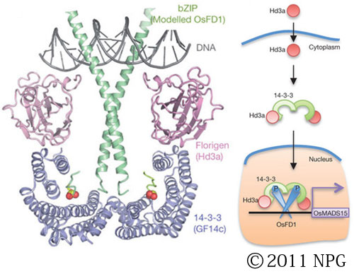 −‘Florigen’ was proposed 75 years ago to be synthesized in the leaf and transported to the shoot apex, where it induces flowering. Only recently have genetic and biochemical studies established that florigen is encoded by FLOWERING LOCUS T (FT), a gene that is universally conserved in higher plants. Nonetheless, the exact function of florigen during floral induction remains poorly understood and receptors for florigen have not been identified. Ko Shimamoto at NAIST and his colleagues have shown that the rice FT homologue Hd3a interacts with 14-3-3 proteins in the apical cells of shoots, yielding a complex that translocates to the nucleus and binds to the Oryza sativa (Os)FD1 transcription factor. The resultant ternary ‘florigen activation complex’ (FAC) induces transcription of OsMADS15, a homologue of A. thaliana APETALA1 (AP1), which leads to flowering. They have determined the crystal structure of rice FAC, which provides a mechanistic basis for florigen function in flowering. Their results indicate that 14-3-3 proteins act as intracellular receptors for florigen in shoot apical cells, and offer new approaches to manipulate flowering in various crops and trees.
| Food / Environment B6 | |
| Structure and function of environmentally-responsive proteins TP Atlas |
|
| Ko Shimamoto | |
| Press Release (in Japanes) from NAIST | |
| Nature, 2011 July 31 14-3-3 proteins act as intracellular receptors for rice Hd3a florigen Ken-ichiro Taoka, Izuru Ohki, Hiroyuki Tsuji, Kyoko Furuita, Kokoro Hayashi, Tomoko Yanase, Midori Yamaguchi, Chika Nakashima, Yekti Asih Purwestri, Shojiro Tamaki, Yuka Ogaki, Chihiro Shimada, Atsushi Nakagawa, Chojiro Kojima& Ko Shimamoto PDB ID code: 3AXY |
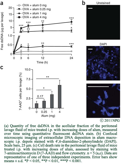 -Aluminum-based adjuvants (aluminum salts or alum) are widely used in human vaccination, although their mechanisms of action are poorly understood. Ken Ishii and his colleagues in Osaka U. and NIBIO in collaboration with researchers at U. Liège in Belgium report that, in mice, alum causes cell death and the subsequent release of host cell DNA, which acts as a potent endogenous immunostimulatory signal mediating alum adjuvant activity. Furthermore, they propose that host DNA signaling differentially regulates IgE and IgG1 production after alum-adjuvanted immunization. They suggest that, on the one hand, host DNA induces primary B cell responses, including IgG1 production, through interferon response factor 3 (Irf3)-independent mechanisms. On the other hand, they suggest that host DNA also stimulates 'canonical' T helper type 2 (TH2) responses, associated with IgE isotype switching and peripheral effector responses, through Irf3-dependent mechanisms. The finding that host cell DNA released from dying cells acts as a damage-associated molecular pattern that mediates alum adjuvant activity may increase their understanding of the mechanisms of action of current vaccines and help in the design of new adjuvants.
| Medicine / Pharmacology A1 | |
| Structural analysis of molecules related to the innate immune system | |
| Ken Ishii | |
| Press Release from Osaka U.; "DNA released from dying host cells mediates aluminum adjuvant activity" Press Release from U. Liège | |
| Nature Medicine, 2011 July 17. DNA released from dying host cells mediates aluminum adjuvant activity Thomas Marichal, Keiichi Ohata, Denis Bedoret, Claire Mesnil, Catherine Sabatel, Kouji Kobiyama, Pierre Lekeux, Cevayir Coban, Shizuo Akira, Ken J Ishii, Fabrice Bureau& Christophe J Desmet |
 -Acetabularia rhodopsin (AR) is a rhodopsin from the marine plant, Acetabularia acetabulum. Shigeyuki Yokoyama at RIKEN and his colleagues reported on their functional and structural analyses of AR. The opsin-encoding gene from A. acetabulum, ARII, was cloned and found to be novel, but homologous to that reported previously. ARII is a light-driven proton pump, as demonstrated by the existence of a photo-induced current through Xenopus oocytes expressing ARII. The photochemical reaction of ARII, prepared by cell-free protein synthesis, was similar to that of bacteriorhodopsin (BR), except for the lack of light-dark adaptation and the different proton release and uptake sequence. The crystal structure, determined at 3.2-A resolution, is the first structure of a eukaryotic member of the microbial rhodopsin family.
| Protein Production C1 | |
| Development of Advanced Production Technologies for Target Proteins | |
| Shigeyuki Yokoyama | |
| Press Release (in Japanese) from RIKEN | |
| J Mol. Biol., 2011 June 25. Crystal structure of the eukaryotic light-driven proton pumping rhodopsin, Acetabularia rhodopsin II, from marine alga Takashi Wada, Kazumi Shimono, Takashi Kikukawa, Masakatsu Hato, Naoko Shinya, So Young Kim, Tomomi Kimura-Someya, Mikako Shirouzu, Jun Tamogami, Seiji Miyauchi, Kwang-Hwan Jung, Naoki Kamo, Shigeyuki Yokoyama. PDB ID code: 3AM6 |
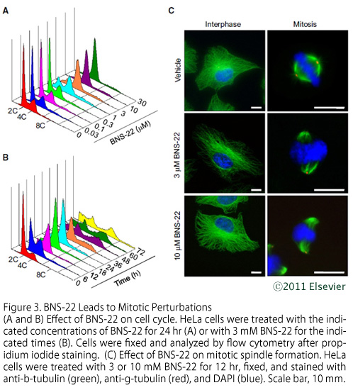 -BNS-22, a chemically synthesized derivative of the natural plant product GUT-70 isolated from the stem bark of Calophyllum brasiliense in Brazil, has antiproliferative activity against human cancer cells, the mechanism of which is unknown. Hiroyuki Osada and his colleagues identified a target of BNS-22 by proteomic profiling analysis, which suggests that BNS-22 belongs to the same cluster as ICRF-193, a DNA topoisomerase II (TOP2) catalytic inhibitor. BNS-22 inhibits kinetoplast DNA decatenation that is mediated by human TOP2A and TOP2B in vitro at an IC50 of 2.8 and 0.42 μM, respectively. BNS-22 does not affect DNA damage and antagonizes TOP2 poison-mediated DNA damage. Like ICRF-193, BNS-22 induces mitotic abnormalities, characterized by impairments in chromosome alignment and segregation, thereby causing polyploidy in HeLa cells. These results indicate that BNS-22 targets TOP2 and acts as its catalytic inhibitor.
| Chemical Regulation C1 | |
| Establishment of Chemical Library and Development of Protein Regulation Technology | |
| Tetsuo Nagano | |
| Press Release (in Japanese) from RIKEN | |
| Chemistry & Biology, 2011 June 24. Identification of a Small-Molecule Inhibitor of DNA Topoisomerase II by Proteomic Profiling. Makoto Kawatani, Hiroshi Takayama, Makoto Muroi, Shinya Kimura, Taira Maekawa, and Hiroyuki Osada. |
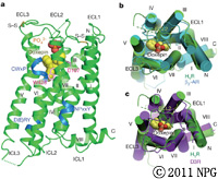 -Histamine is an important pharmacological mediator involved in pathophysiological processes such as allergies and inflammations. Histamine H1 receptor (H1R) antagonists are very effective drugs alleviating the symptoms of allergic reactions. An international team led by So Iwata of Kyoto U. showed the crystal structure of the H1R complex with doxepin, a first-generation H1R antagonist. Doxepin sits deep in the ligand-binding pocket and directly interacts with Trp?428, a highly conserved key residue in G-protein-coupled-receptor activation. This well-conserved pocket with mostly hydrophobic nature contributes to the low selectivity of the first-generation compounds. The pocket is associated with an anion-binding region occupied by a phosphate ion. Docking simulation of various second-generation H1R antagonists reveals that the unique carboxyl group present in this class of compounds interacts with Lys?191and/or Lys?179, both of which form part of the anion-binding region. This region is not conserved in other aminergic receptors, demonstrating how minor differences in receptors lead to pronounced selectivity differences with small molecules. Their study sheds light on the molecular basis of H1R antagonist specificity against H1R.
| Fundamental Biology B4 | |
| Membrane transporters: structure and function of important drug targets TP Atlas |
|
| So Iwata | |
| Press Release (in Japanese) from JST - Kyoto U. - Kyushu U. Press Release (in English) from Diamond Light Source |
|
| Nature, 2011 June22. Structure of the human histamine H1receptor complex with doxepin. Tatsuro Shimamura, Mitsunori Shiroishi, SimoneWeyand, HirokazuTsujimoto,GraemeWinter,VsevolodKatritch, Ruben Abagyan, Vadim Cherezov,Wei Liu, GyeWon Han, Takuya Kobayashi, Raymond C. Stevens & So Iwata. PDB ID code: 3RZE |
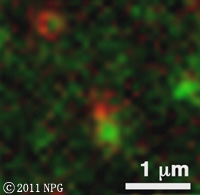 -Endosomal trafficking plays an integral role in various eukaryotic cell activities and serves as a basis for higher-order functions in multicellular organisms. However, the molecular mechanism of endosomal trafficking in plants is mostly unknown. The identification of lineage-specific regulators in eukaryotes indicates that endosomal trafficking is diversified according to distinct body plans and lifestyles. In addition to orthologues of metazoan RAB5, land plants possess a unique RAB5 molecule, which is one of the most prominent features of plant RAB GTPase organization. Plants have also evolved a unique repertoire of SNAREs, the most distinctive of which are diverse VAMP7-related longins, including plant-unique VAMP72 derivatives. Takashi Ueda and his colleagues demonstrate that a plant-unique RAB5 protein, ARA6, acts in an endosomal trafficking pathway in Arabidopsis thaliana. ARA6 modulates the assembly of a distinct SNARE complex from conventional RAB5, and has a functional role in the salinity stress response. Their results indicate that plants possess a unique endosomal trafficking network and provide the first indication of a functional link between a specific RAB and a specific SNARE complex in plants.
| Fundamental Biology B6 | |
| Structure-function analysis of protein complexes that regulate vesicular traffic TP Atlas |
|
| Soichi Wakatsuki | |
| Press Release from U. Tokyo; "How did plants explore unique intracellular trafficking routes?" | |
| Nature Cell Biology, 2011 June12. A membrane trafficking pathway regulated by the plant-specific RAB GTPase ARA6. Kazuo Ebine, Masaru Fujimoto, Yusuke Okatani, Tomoaki Nishiyama, Tatsuaki Goh, Emi Ito, Tomoko Dainobu, Aiko Nishitani, Tomohiro Uemura, Masa H. Sato, Hans Thordal-Christensen, Nobuhiro Tsutsumi, Akihiko Nakano and Takashi Ueda. |
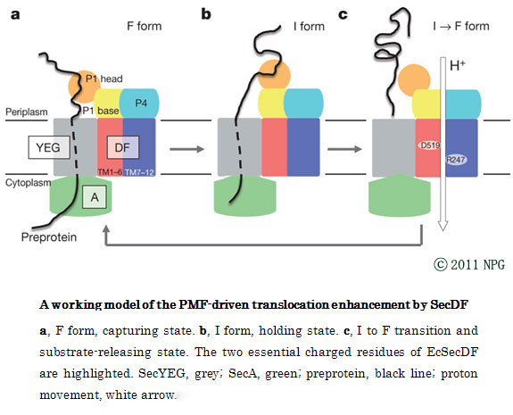 -Protein translocation across the bacterial membrane, mediated by the secretory translocon SecYEG and the SecA ATPase, is enhanced by proton motive force and membrane-integrated SecDF, which associates with SecYEG. The role of SecDF has remained unclear, although it is proposed to function in later stages of translocation. Osamu Nureki and his colleagues determined the crystal structure of Thermus thermophilus SecDF at 3.3Å resolution. Higher-resolution analysis of the periplasmic domains suggested that the major periplasmic domain P1, which binds an unfolded protein, undergoes functionally important conformational changes. In vitro analyses identified an ATP-independent step of protein translocation that requires both SecDF and proton motive force. Electrophysiological analyses revealed that SecDF conducts protons in a manner dependent on pH and the presence of an unfolded protein. Therefore, they propose that SecDF functions as a membrane integrated chaperone, powered by proton motive force, to achieve ATP-independent protein translocation.
| Fundamental Biology B5 | |
| Elucidation of the mechanism of high-order cellular functions achieved by non-coding RNAs | |
| Osamu Nureki | |
| Press Release from U. Tokyo - Kyoto U. - Kyoto Sangyo U.; | |
| Nature, 2011 May12. Structure and function of a membrane component SecDF that enhances protein export. Tomoya Tsukazaki, Hiroyuki Mori, Yuka Echizen, Ryuichiro Ishitani, Shuya Fukai Takeshi Tanaka, Anna Perederina, Dmitry G. Vassylyev, Toshiyuki Kohno, Andre's D. Maturana, Koreaki Ito & Osamu Nureki. Graduate School of Life and Environmental Sciences, University of Tsukuba, Tsukuba, Ibaraki 305-8577, Japan. PDB ID code: 3AQP, 3AQO, 2RRN |
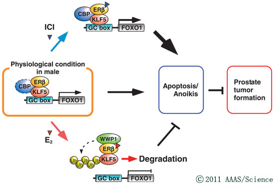 -Clinical evidence suggests that antiestrogens inhibit the development of androgen-insensitive prostate cancer. Junn Yanagisawa and his colleagues at U. Tsukuba, U.Tokyo and Tohoku U. revealed that the estrogen receptor β (ERβ) mediates inhibition by the antiestrogen ICI 182,780 (ICI) and its enhancement by estrogen. ERβ associated with gene promoters through the tumor-suppressing transcription factor KLF5. ICI treatment increased the recruitment of the transcription coactivator CBP to the promoter of FOXO1 through ERβ and KLF5, which enhanced the transcription of FOXO1. The increase in FOXO1 abundance led to anoikis in prostate cancer cells, thereby suppressing tumor growth. In contrast, estrogen induced the formation of complexes containing ERβ, KLF5, and the ubiquitin ligase WWP1, resulting in the ubiquitination and degradation of KLF5. The combined presence of KLF5 and ERβ positively correlated with longer cancer-specific survival in prostate cancer patients. Our results demonstrate that estrogens and antiestrogens affect prostate tumor growth through ERβ-mediated regulation of KLF5. The development of ERβ-based therapies may lead to improved drug efficacy.
| Medicine / Pharmacology B2 | |
| Development of new compounds for fibrosis therapy | |
| Junn Yanagisawa | |
| Press Release (in Japanese) from U. Tsukuba; | |
| Sci. Signal. 2011 April 12. Estrogen Regulates Tumor Growth Through a Nonclassical Pathway that Includes the Transcription Factors ERβ and KLF5. Y. Nakajima, K. Akaogi, T. Suzuki, A. Osakabe, C. Yamaguchi, N. Sunahara, J. Ishida, K. Kako, S. Ogawa, T. Fujimura, Y. Homma, A. Fukamizu, A. Murayama, K. Kimura, S. Inoue, J. Yanagisawa. Graduate School of Life and Environmental Sciences, University of Tsukuba, Tsukuba, Ibaraki 305-8577, Japan. |
|
| Sci. Signal. 2011 April 12. Estrogen Receptor β: Switching to a New Partner and Escaping from Estrogen. Y.-K. Leung, S.-M. Ho. |
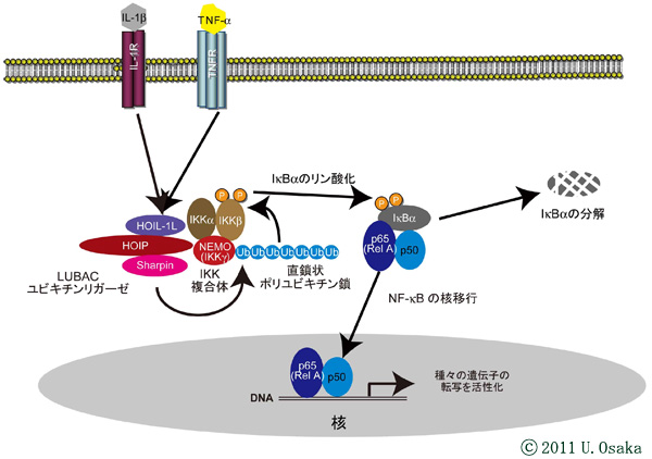 -NF-κB activated by various stimuli is a transcription factor involved in various functions, including inflammation and cell survival. Researchers led by Kazuhiro Iwai had previously reported that linear ubiquitin chain assembly complex (LUBAC) induces NF-κB activation through conjugation of linear polyubiquitin chains to NEMO and that LUBAC is composed of the two proteins HOIL-1L and HOIP. Now, they identify SHARPIN as an additional component of LUBAC. SHARPIN-containing complexes can linearly ubiquitinate NEMO and activated NF-κB. Spontaneous null mutations in the SHARPIN gene are responsible for the cpdm (chronic proliferative dermatitis) phenotype in mice. Deletion of SHARPIN drastically reduced the amount of LUBAC, which resulted in attenuated TNF-α- and CD40-mediated activation of NF-κB in mouse embryonic fibroblasts or B cells from cpdm mice. These results strongly suggest the involvement of LUBAC-induced NF-κB activation in various disorders.
| Fundamental Biology A8 | |
| Mechanism underlying selective activation of NF-κB via linear polyubiquitination | |
| Kazuhiro Iwai | |
| Press Release from Osaka U.; "SHARPIN is a component of the NF-κB-activating linear ubiquitin chain assembly complex " | |
| Nature., 2011 March 31. SHARPIN is a component of the NF-κB-activating linear ubiquitin chain assembly complex. Fuminori Tokunaga, Tomoko Nakagawa, Masaki Nakahara, Yasushi Saeki, Masami Taniguchi, Shin-ichi Sakata, Keiji Tanaka, Hiroyasu Nakano & Kazuhiro Iwai. Department of Biophysics and Biochemistry, Graduate School of Medicine, Osaka University, Suita, Osaka 565-0871, Japan. |
 -One of the key factors concerning the preparation of membrane protein samples for structural and functional studies is whether the proteins retain their correct physiological folding and oligomerization states. Membrane proteins often lose their native folding or oligomerization during sample preparation. Dr. Atsuko Yamashita and her colleagues at RIKEN reported an improved native polyacrylamide gel electrophoresis (PAGE) method capable of evaluating the hydrodynamic states of membrane proteins and allowing in-gel fluorescence detection. In this method, bis(alkyl)sulfosuccinate is used to provide negative charges for detergent-solubilized membrane proteins to facilitate proper electrophoretic migration without disturbing their native hydrodynamic states. The method achieved high-resolution electrophoretic separation, in good agreement with the elution profiles obtained by size exclusion chromatography. The applicability of in-gel fluorescence detection for tagged green fluorescent protein (GFP) facilitates the analysis of samples without any purification. This method might serve as a general analytical technique for assessing the folding, oligomerization, and protein complex formation of membrane proteins.
| Protein Production D2 | |
| Development of innovative methods to support membrane protein crystallization | |
| Hiroaki Kato | |
| Press Release (in Japanese) from RIKEN; | |
| Anal Biochem., 2011 Feb 1. High-resolution native polyacrylamide gel electrophoresis for membrane proteins capable of fluorescence detection and hydrodynamic state evaluation. Ihara M, Matsuura N, Yamashita A. Molecular Signaling Research Team, Structural Physiology Research Group, RIKEN SPring-8 Center, Kouto, Sayo, Hyogo 679-5148, Japan. |
 -The proteins that form the bacterial flagellum are translocated to its distal end through the central channel of the growing flagellum by the flagellar-specific protein export apparatus, a family of the type III protein secretion system. FliI and FliJ are soluble components of this apparatus. FliI is an ATPase that has extensive structural similarity to theαand β subunits of FoF1-ATP synthase. FliJ is essential for export, but its function remains obscure. Katsumi Imada of Osaka U. and his colleagues revealed that the structure of FliJ is remarkably similar to that of the two-stranded α-helical coiled-coil part of the γ subunit of FoF1-ATP synthase and that FliJ promotes the formation of FliI hexamer rings by binding to the center of the ring. These results suggest that the type III protein export system and F- and V-type ATPases share a similar mechanism and an evolutionary relationship.
| Fundamental Biology A1 | |
| Structural and functional studies of bacterial type Ⅲ and type | |
| Katsumi Imada | |
| Press Release (in Japanese) from the Osaka University; | |
| Nat Struct Mol Biol., 2011 Jan 30. Common architecture of the flagellar type III protein export apparatus and F- and V-type ATPases. Ibuki T, Imada K, Minamino T, Kato T, Miyata T, Namba K. Graduate School of Frontier Biosciences, Osaka University, Osaka, Japan. PDB ID code: 3AJW |
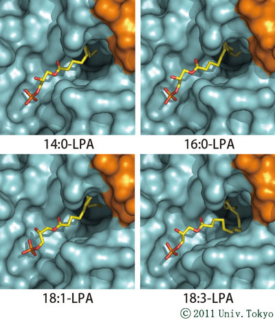 -NPP2 (also known as Autotaxin) is a secreted lysophospholipase D that hydrolyzes lysophosphatidylcholine to generate lysophosphatidic acid (LPA), a lipid mediator that activates G protein-coupled receptors to evoke various cellular responses. Osamu Nureki, Junken Aoki, Junichi Takagi and their colleagues have reported the crystal structures of mouse NPP2 alone and in complex with LPAs with different acyl-chain lengths and saturations. These structures reveal that the multidomain architecture helps to maintain the structural rigidity of the lipid-binding pocket, which accommodates the respective LPA molecules in distinct conformations. They indicate that a loop region in the catalytic domain is a major determinant for the substrate specificity of the NPP family enzymes. Furthermore, along with biochemical and biological data, these structures suggest that the produced LPAs are delivered from the active site to cognate G proteincoupled receptors through a hydrophobic channel.
| Medicine/Pharmacology B3 | |
| Structure and drug development of NPP family members involved in cancer and various diseases | |
| Junken Aoki | |
| News Release from the University of Tokyo; "Structure of a protein associated with cancer metastasis" | |
| Nature Structural & Molecular Biology, January 16, 2011. Crystal structure of autotaxin and insight into GPCR activation by lipid mediators. Hiroshi Nishimasu, Shinichi Okudaira, Kotaro Hama, Emiko Mihara, Naoshi Dohmae, Asuka Inoue, Ryuichiro Ishitani, Junichi Takagi, Junken Aoki& Osamu Nureki. Department of Biophysics and Biochemistry, Graduate School of Science, The University of Tokyo, Tokyo, Japan. PDB ID: 3NKM, 3NKN, 3NKO, 3NKP, 3NKQ, 3NKR |
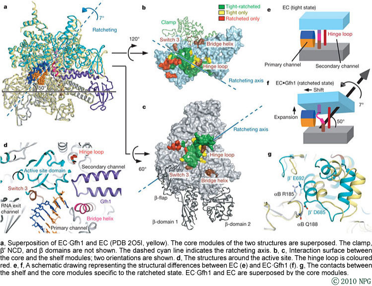 -The RNA polymerase (RNAP) is the principal enzyme of transcription for gene expression. Transcription is regulated by various transcription factors. Gre factor homologue 1 (Gfh1), found in the Thermus genus, inhibits transcription initiation and elongation by binding directly to RNAP. Researchers led by Shigeyuki Yokoyama at the Univ. Tokyo report the crystal structure of Thermus thermophilus RNAP complexed with Gfh1. The amino-terminal domain of Gfh1 fully occludes the channel formed between the two central modules of RNAP; this channel would normally be used for nucleotide triphosphate (NTP) entry into the catalytic site. The NTP-entry channel is expanded, because the central modules are ‘ratcheted’, as compared with the previously reported elongation complexes. Simultaneously, the ratcheting expands the nucleic-acid-binding channel, and kinks the bridge helix. The proposed molecular mechanisms of transcription inhibition by preventing NTP binding to bacterial RNAP could lead to developments of novel antibiotics.
| Protein Production C1 | |
| Development of advanced production technologies for target proteins | |
| Shigeyuki Yokoyama | |
| News Release from the University of Tokyo; "A novel mechanism to inhibit bacterial gene expression - A spiky transcription factor sticks and stops RNA polymerase -" | |
| Nature, December 1, 2010. Crystal structure of bacterial RNA polymerase bound with a transcription inhibitor protein. Shunsuke Tagami, Shun-ichi Sekine, Thirumananseri Kumarevel, Nobumasa Hino, Yuko Murayama, Syunsuke Kamegamori, Masaki Yamamoto, Kensaku Sakamoto & Shigeyuki Yokoyama. Department of Biophysics and Biochemistry, Graduate School of Science, University of Tokyo, 7-3-1 Hongo, Bunkyo-ku, Tokyo 113-0033, Japan. PDB ID code: 3AOH, 3AOI |
 -Semaphorins and their receptor plexins constitute a cell-signaling system used in various biological processes, and both protein families have been implicated in numerous diseases. The binding of semaphorins to the plexin ectodomain activates plexin’s intrinsic GTPase-activating protein (GAP) at the cytoplasmic region, ultimately modulating cellular adhesion behavior. Researchers led by Junichi Takagi of Osaka U. reported the crystal structures of the semaphorin 6A (Sema6A) receptor-binding fragment and the plexin A2 (PlxnA2) ligand-binding fragment in both their pre-signaling and signaling states. Before binding, the Sema6A ectodomain was in the expected ‘face-to-face’ homodimer arrangement, whereas PlxnA2 was in an unexpected ‘head-on’ homodimer arrangement. In contrast, the structure of the Sema6A?PlxnA2 signaling complex revealed a 2:2 heterotetramer in which the two PlxnA2 monomers dissociated from one another and docked onto the top face of the Sema6A homodimer using the same interface as the head-on homodimer, indicating that plexins undergo ‘partner exchange’. Thus, homodimer-to-heterodimer transitions of cell-surface plexin that result in a specific orientation of its molecular axis relative to the membrane may constitute the structural mechanism by which the ligand-binding ‘signal’ is transmitted to the cytoplasmic region, inducing GAP domain rearrangements and activation.
| Protein Production D1: Development of novel affinity tag system for the high-quality production of extracellular and membrane proteins(Principal Investigator: Junichi Takagi) Medicine/Pharmacology B4: Structural and functional analysis of semaphorins and their receptors(Principal Investigator: Atsushi Kumanogoh) |
|
| News Release from Osaka University; "Structural basis for semaphorin signalling through the plexin receptor" | |
| Nature, September 30 , 2010. Structural basis for semaphorin signalling through the plexin receptor. Terukazu Nogi, Norihisa Yasui, Emiko Mihara, Yukiko Matsunaga, Masanori Noda, Naoya Yamashita, Toshihiko Toyofuku, Susumu Uchiyama, Yoshio Goshima, Atsushi Kumanogoh & Junichi Takagi. Laboratory of Protein Synthesis and Expression, Institute for Protein Research, Osaka University, Suita, Osaka 565-0871, Japan. PDB ID code: 3AFC, 3AL9, 3AL8 |
 -In most bacteria and all archaea, glutamyl-tRNA synthetase (GluRS) glutamylates both tRNAGlu and tRNAGln, and then Glu-tRNAGln is selectively converted to Gln-tRNAGln by a tRNA-dependent amidotransferase. Shigeyuki Yokoyama and Takuhiro Ito at RIKEN reported the formation of the ‘glutamine transamidosome’ from the bacterium Thermotoga maritima, consisting of tRNAGln, GluRS and the heterotrimeric amidotransferase GatCAB, and its crystal structure. The anticodon-binding body of GluRS recognizes the common features of tRNAGln and tRNAGlu, whereas the tail body of GatCAB recognizes the outer corner of the L-shaped tRNAGln. GluRS is in the productive form, as its catalytic body binds to the amino-acid-acceptor arm of tRNAGln. In contrast, GatCAB is in the non-productive form: the catalytic body of GatCAB contacts that of GluRS and is located near the acceptor stem of tRNAGln, in an appropriate site to wait for the completion of Glu-tRNAGln formation by GluRS. They proposed the mechanism by which the transamidosome efficiently performs the two consecutive steps of Gln-tRNAGln formation, with a low risk of releasing the unstable intermediate Glu-tRNAGln.
| Fundamental Biology B5: Elucidation of the mechanism of high-order cellular functions achieved by non-coding RNAs (Principal Investigator: Osamu Nureki) Protein Production C1: Development of Advanced Production Technologies for Target Proteins (Principal Investigator: Shigeyuki Yokoyama) |
|
| News Release (in Japanese) from RIKEN & University of Tokyo; | |
| Nature, September 30, 2010. Two enzymes bound to one transfer RNA assume alternative conformations for consecutive reactions . Takuhiro Ito & Shigeyuki Yokoyama. Department of Biophysics and Biochemistry, Graduate School of Science, The University of Tokyo, Hongo, Bunkyo-ku, Tokyo 113-0033, Japan. PDB ID code: 3AKZ, 3AL0 |
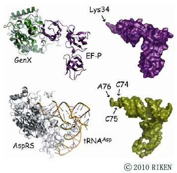 -Aminoacyl-tRNA synthetase (aaRS) paralogs with unknown functions exist in various species. Researchers headed by Shigeyuki Yokoyama at RIKEN reported novel 'protein lysylation' by an Escherichia coli lysyl-tRNA synthetase paralog, GenX. X-ray crystallographic analysis showed that the structure of the GenX protein resembles that of a class II aaRS. Further in vitro studies revealed that it specifically aminoacylates the translation elongation factor P (EF-P) with lysine. The shape of the protein substrate mimics that of the L-shaped tRNA, and its lysylation site corresponds to the tRNA 3′ end. Thus, they showed how the aaRS architecture can be adapted to achieve aminoacylation of a specific protein. Moreover, in vivo analyses revealed that EF-P lysylation by GenX is enhanced by YjeK (lysine 2,3-aminomutase paralog), which is encoded next to the EF-P gene, and might convert α-lysyl?EF-P to β-lysyl?EF-P. In vivo analyses indicated that the EF-P modification by GenX and YjeK is essential for cell survival.
| Fundamental Biology B5 | |
| Elucidation of the mechanism of high-order cellular functions achieved by non-coding RNAs | |
| Osamu Nureki | |
| Research Highlight from RIKEN; "Copycat protein finds a perfect match" |
|
| Nature Struct. Molec. Biol., August 22, 2010. A paralog of lysyl-tRNA synthetase aminoacylates a conserved lysine residue in translation elongation factor P. Tatsuo Yanagisawa, Tomomi Sumida, Ryohei Ishii, Chie Takemoto & Shigeyuki Yokoyama. RIKEN Systems and Structural Biology Center, Tsurumi, Yokohama, Japan. PDB ID code: 3A5Y, 3A5Z |
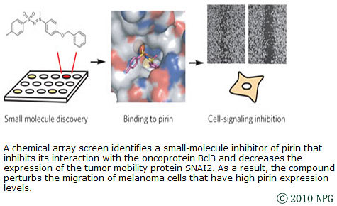 -The discovery of small molecules that bind to a specific target and disrupt the function of proteins is an important step in chemical biology, especially for poorly characterized proteins. Human pirin is a nuclear protein of unknown function that is widely expressed in punctate subnuclear structures in human tissues. Researchers led by Hiroyuki Osada at RIKEN report the discovery of a small molecule that binds to pirin. They determined how the small molecule bound to pirin by solving the cocrystal structure. Either knockdown of pirin or treatment with the small molecule inhibited melanoma cell migration. Thus, inhibition of pirin by the small molecule has led to a greater understanding of the function of pirin and represents a new method of studying pirin-mediated signaling pathways.
| Chemical Regulation C1 | |
| Establishment of Chemical Library and Development of Protein Regulation Technologys | |
| Tetsuo Ngano | |
| News Release (in English) from RIKEN; "Small-molecule inhibitor uncovers protein role in melanoma cell migration" |
|
| Nature Chemical Biology, August 15, 2010. A small-molecule inhibitor shows that pirin regulates migration of melanoma cells. Isao Miyazaki, Siro Simizu, Hideo Okumura, Satoshi Takagi & Hiroyuki Osada. Chemical Library Validation Team, Chemical Biology Core Facility, Chemical Biology Department, RIKEN Advanced Science Institute, Wako, Saitama, Japan. PDB ID code: 3ACL |
 -The 21st amino acid, selenocysteine (Sec), is assigned to the codon UGA and is biosynthesized on the Sec-specific tRNA (tRNASec). In archaea and eukarya, tRNASec is ligated with serine by seryl-tRNA synthetase (SerRS), the seryl moiety is phosphorylated by O-phosphoseryl-tRNA kinase (PSTK), and the phosphate group is replaced with selenol by Sep-tRNA:Sec-tRNA synthase. PSTK selectively phosphorylates seryl-tRNASec, whereas SerRS ligates serine with both serine-specific tRNA (tRNASer) and tRNASec. Researchers headed by Shigeyuki Yokoyama at RIKEN determined the crystal structures of the tRNASec・PSTK complex. The structures revealed that the small C-terminal domain (CTD) of the PSTK is associated with the characteristic D arms of tRNASec, independently of the N-terminal catalytic domain. The specific interaction between the unique tRNASec D arm and the PSTK CTD accounts for the strict tRNASec selectivity of PSTK and guarantees the UGA-specific encoding of selenocysteine.
| Fundamental Biology B5 | |
| Elucidation of the mechanism of high-order cellular functions achieved by non-coding RNAs | |
| Osamu Nureki | |
| News Release (in English) from RIKEN & University of Tokyo; "Scientists clarify structural basis for biosynthesis of mysterious 21st amino acid" |
|
| Molecular Cell, August 13, 2010. Structural Basis for the Major Role of O-Phosphoseryl-tRNA Kinase in the UGA-Specific Encoding of Selenocysteine. Chiba S, Itoh Y, Sekine SI, Yokoyama S. Department of Biophysics and Biochemistry, Graduate School of Science, The University of Tokyo, 7-3-1 Hongo, Bunkyo-ku, Tokyo, Japan. PDB ID code: 3ADB, 3ADC, 3ADD |
 -Although C-nitroso aromatic compounds have several bioactivities of medicinal interest, the biosynthetic enzymes involved in C-nitrosation have remained unknown until now. Researchers led by Yasuo Ohnishi at the University of Tokyo report the entire biosynthesis pathway of 4-hydroxy-3-nitrosobenzamide in Streptomyces murayamaensis, in which a tyrosinase-like copper-containing monooxygenase is responsible for the C-nitrosation. This finding indicates diverse catalytic functions of tyrosinase-like copper-containing monooxygenases in nature.
| Food/Environment A2 | |
| Structure-based functional analysis of key enzymes that can be applied to production of antibiotics and other useful compounds | |
| Yasuo Ohnishi | |
| News Release (in Japanese) from University of Tokyo; | |
| Nature Chemical Biology, August 1, 2010. A copper-containing oxidase catalyzes C-nitrosation in nitrosobenzamide biosynthesis. Akio Noguchi, Takeshi Kitamura, Hiroyasu Onaka, Sueharu Horinouchi & Yasuo Ohnishi. Department of Biotechnology, Graduate School of Agriculture and Life Sciences, University of Tokyo, Tokyo, Japan. |
 -Various social behaviors in mice are regulated by chemical signals called pheromones that act through the vomeronasal system. Exocrine gland-secreting peptide 1 (ESP1) is a 7-kDa peptide that is released into male tear fluids and stimulates vomeronasal sensory neurons in female mice. Kazushige Touhara at the University of Tokyo and his colleagues describe the molecular and neural mechanisms that are involved in the decoding of ESP1 signals in the vomeronasal system, which leads to behavioral output in female mice. ESP1 is recognized by a specific vomeronasal receptor, V2Rp5, and the ligand?receptor interaction results in sex-specific signal transmission to the amygdaloid and hypothalamic nuclei via the accessory olfactory bulb. Consequently, ESP1 enhances female sexual receptive behavior upon male mounting (lordosis), allowing successful copulation. In V2Rp5-deficient mice, ESP1 induces neither neural activation nor sexual behavior. These findings show that ESP1 is a crucial male pheromone that regulates female reproductive behavior through a specific receptor in the mouse vomeronasal system.
| Food/Environment B1 | |
| Structural and functional analyses of the rodent ESP family | |
| Hiroaki Terasawa | |
| News Release (in Japanese) from University of Tokyo; | |
| ScienceNow | |
| Nature, July 1, 2010. The male mouse pheromone ESP1 enhances female sexual receptive behaviour through a specific vomeronasal receptor. Sachiko Haga, Tatsuya Hattori, Toru Sato, Koji Sato, Soichiro Matsuda, Reiko Kobayakawa, Hitoshi Sakano, Yoshihiro Yoshihara, Takefumi Kikusui and Kazushige Touhara. Departmentof Applied Biological Chemistry, Graduate School of Agricultural and Life Sciences, The University of Tokyo, Tokyo 113-8657, Japan. |
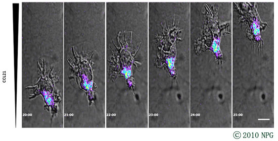 -The recirculation of leukocytes is essential for proper immune responses. However, the molecular mechanisms that regulate the entry of leukocytes into the lymphatics remain unclear. Atsushi Kumanogoh at Osaka University and his colleagues show that plexin-A1, a principal receptor component for class III and class VI semaphorins, was crucially involved in the entry of dendritic cells (DCs) into the lymphatics. Additionally, they show that the semaphorin Sema3A was required for DC transmigration and that Sema3A produced by the lymphatics promoted actomyosin contraction at the trailing edge of migrating DCs. Their findings not only demonstrate that semaphorin signals are involved in DC trafficking but also identify a previously unknown mechanism that induces actomyosin contraction as these cells pass through narrow gaps.
| Medicine/Pharmacology B4 | |
| Structural and functional analysis of semaphorins and their receptors | |
| Atsushi Kumanogoh | |
| News Release from Osaka University; "Semaphorins guide the entry of dendritic cells into the lymphatics by activating myosin II" | |
| Nature Immunology, May 30, 2010. Semaphorins guide the entry of dendritic cells into the lymphatics by activating myosin II. Hyota Takamatsu, Noriko Takegahara, Yukinobu Nakagawa, Michio Tomura, Masahiko Taniguchi, Roland H Friedel, Helen Rayburn, Marc Tessier-Lavigne, Yutaka Yoshida, Tatsusada Okuno, Masayuki Mizui, Sujin Kang, Satoshi Nojima, Tohru Tsujimura, Yuji Nakatsuji, Ichiro Katayama, Toshihiko Toyofuku, Hitoshi Kikutani & Atsushi Kumanogoh. Department of Immunopathology, Research Institute for Microbial Diseases, Osaka University, Osaka, Japan. |
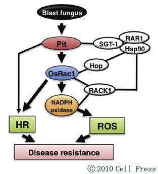 -Ko Shimamoto and his colleagues found that the rice GTPase OsRac1 at the plasma membrane interacts directly with Pit, an NLR protein that confers resistance to the rice blast fungus. OsRac1 contributes to Pit-mediated ROS (reactive oxygen species) production as well as the hypersensitive response and is required for Pit-mediated disease resistance in rice. Furthermore, the active form of Pit induces the activation of OsRac1 at the plasma membrane. Thus, OsRac1 is activated by Pit during pathogen attack and plays a critical role in Pit-mediated immunity in rice.
| Food/Environment B6 | |
| Structure and function of environmentally-responsive proteins | |
| Ko Shimamoto | |
| News Release (in Japanese) from NAIST; | |
| Cell Host Microbe., May 20, 2010. Activation of a Rac GTPase by the NLR Family Disease Resistance Protein Pit Plays a Critical Role in Rice Innate Immunity. Kawano Y, Akamatsu A, Hayashi K, Housen Y, Okuda J, Yao A, Nakashima A, Takahashi H, Yoshida H, Wong HL, Kawasaki T, Shimamoto K. Laboratory of Plant Molecular Genetics, Nara Institute of Science and Technology, 8916-5 Takayama, Ikoma, Nara 630-0101, Japan. |
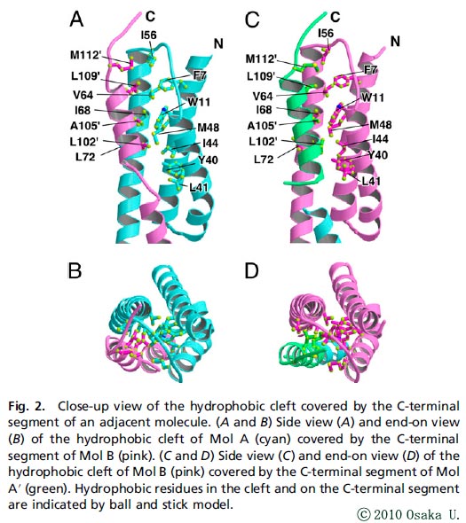 -For self-assembly of the bacterial flagellum, the flagellar component proteins synthesized in the cytoplasm are exported by the flagellar type III export apparatus to the growing, distal end. Flagellar protein export is highly organized and well controlled. Flagellar-specific chaperones not only facilitate the export of their cognate proteins, as well as prevent their premature aggregation, but also play a role in fine tuning flagellar gene expression to be coupled with the flagellar assembly process. FliT is a chaperone responsible for the export of the filament-capping protein FliD and for negative control of flagellar gene expression by binding to the FlhDC complex. Katsumi Imada and his colleagues at Osaka U. reported the crystal structure of Salmonella FliT. The structural and biochemical analyses reveal that the C-terminal segment of FliT regulates its interactions with the FlhDC complex, FliI, and FliJ, and that its conformational change is responsible for the switch in its binding partners during flagellar protein export.
| Fundamental Biology A1 | |
| Structural and functional studies of bacterial type Ⅲ and type Ⅳ protein export systems | |
| Katsumi Imada | |
| News Release (in Japanese) from Osaka University; | |
| Proc Natl Acad Sci USA., April 26, 2010. Structural insight into the regulatory mechanisms of interactions of the flagellar type III chaperone FliT with its binding partners. Imada K, Minamino T, Kinoshita M, Furukawa Y, Namba K. Graduate School of Frontier Biosciences, Osaka University, Suita, Osaka 565-0871, Japan. PDB: 3A7M |
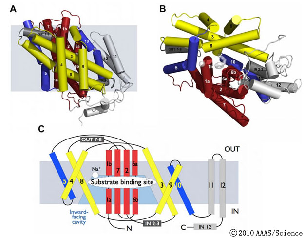 -Hydantoin membrane transporter (Mhp1) mediates the uptake of hydantoins as part of a metabolic salvage pathway for their conversion to amino acids. Researchers led by So Iwata of Kyoto U. already solved the Mhp1 structures of the outward-facing open and substrate-bound occluded conformations. Now, they report the crystal structure of an inward-facing conformation. From analyses of the three structures and molecular dynamics simulations, they proposed a mechanism for the transport cycle in Mhp1, providing the basis of an alternating access mechanism applicable to many transporters of the emerging superfamily.
| Fundamental Biology B4 | |
| Membrane transporters: structure and function of important drug targets | |
| So Iwata | |
| News Release (in Japanese) from JST - Kyoto U. News Release from U. Leeds; |
|
| Science, April 23, 2010. Molecular Basis of Alternating Access Membrane Transport by the Sodium-Hydantoin Transporter Mhp1. Tatsuro Shimamura, Simone Weyand, Oliver Beckstein, Nicholas G. Rutherford, Jonathan M. Hadden, David Sharples, Mark S. P. Sansom, So Iwata, Peter J. F. Henderson, and Alexander D. Cameron. Department of Cell Biology, Graduate School of Medicine, Kyoto University, Yoshida-Konoe-cho, Sakyo-Ku, Kyoto 606-8501, Japan. PDB ID code: 2X79 |
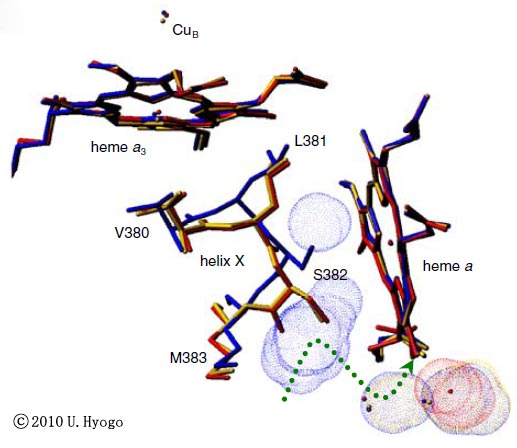 -Cytochrome c oxidase is the terminal oxidase of cellular respiration, which reductively converts molecular oxygen (O2) to two water molecules coupled to a proton-pumping process. Researchers headed by Shinya Yoshikawa at U. Hyogo probed the O2 reduction site of cytochrome c oxidase by x-ray structural analyses of CO, NO, and CN- derivatives to investigate the mechanism of the complete reduction of O2. Their investigations identify critical roles for the O2 reduction site in stabilizing the O2-bound form, facilitating a complete reduction of O2 with minimal production of reactive oxygen species, and providing a gate for effective proton pumping.
| Fundamental Biology B3 | |
| 3D structural and functional analyses for elucidation of the mechanism of mitochondrial respiration | |
| Shinya Yoshikawa | |
| News Release (in Japanese) from U. Hyogo; | |
| Proc Natl Acad Sci USA, April 12, 2010. Bovine cytochrome c oxidase structures enable O2 reduction with minimization of reactive oxygens and provide a proton-pumping gate. Muramoto K, Ohta K, Shinzawa-Itoh K, Kanda K, Taniguchi M, Nabekura H, Yamashita E, Tsukihara T, Yoshikawa S. Department of Life Science, University of Hyogo, 3-2-1 Kouto, Kamigohri, Akoh, Hyogo 678-1297, Japan. PDB ID code: 3AG1, 3AG2, 3AG3, 3AG4 |
 -Ken Ishii at Osaka Univ. and his colleagues examined the mechanisms of action for the three different types of influenza virus vaccines (live virus, inactivated whole-virus and split-virus) using mice lacking three types of sensor receptors (TLR7, IPS-1 and ASC). They revealed that TLR7 acted as the adjuvant receptor for the immunogenicity of both live virus and whole-virus vaccines, whereas a split vaccine failed to immunize naive mice. Furthermore, whole-virus vaccine loaded plasmacytoid dendritic cells are found to be sufficient to transfer immunogenicity to naive mice, which requires intrinsic as well as extrinsic type I interferon signaling.
| Medicine/Pharmacology A1 | |
| Structural analysis of molecules related to the innate immune system | |
| Shizuo Akira, Ken Ishii | |
| News Release from Osaka University; "Plasmacytoid Dendritic Cells Delineate Immunogenicity of Influenza Vaccine Subtypes" |
|
| Business Week | |
| Science Translational Medicine, March 31, 2010. Plasmacytoid Dendritic Cells Delineate Immunogenicity of Influenza Vaccine Subtypes Shohei Koyama, Taiki Aoshi, Takeshi Tanimoto, Yutaro Kumagai, Kouji Kobiyama, Takahiro Tougan, Kazuo Sakurai, Cevayir Coban, Toshihiro Horii, Shizuo Akira, and Ken J. Ishii. Laboratory of Host Defense, WPI Immunology Frontier Research Center, Osaka University, Suita, Osaka 565-0871, Japan. |
 -Researcher headed by Takashi Kadowaki of the Univ. Tokyo revealed that AdipoR1 has a crucial role in the physiological and pathophysiological significance of adiponectin in muscle, and is involved in mitochondrial function and oxidative stress, glucose and lipid metabolism, and exercise endurance. This study suggests that agonism of AdipoR1, as well as strategies to increase AdipoR1 in muscle, may be logical approaches to providing a new treatment modality for mitochondrial dysfunction, insulin resistance and type 2 diabetes linked to obesity.
| Medicine/Pharmacology A6 | |
| Structure-based functional analyses and development of drug intervention in metabolic syndrome and diabetes- AdipoR/AMPK/ACC as key targets | |
| Takashi Kadowaki | |
| News Release (in Japanese) from the University of Tokyo; | |
| Nature. 2010 Mar 31. [Epub ahead of print] Adiponectin and AdipoR1 regulate PGC-1α and mitochondria by Ca2+ and AMPK/SIRT1 Masato Iwabu, Toshimasa Yamauchi, Miki Okada-Iwabu, Koji Sato, Tatsuro Nakagawa, Masaaki Funata, Mamiko Yamaguchi, Shigeyuki Namiki, Ryo Nakayama, Mitsuhisa Tabata, Hitomi Ogata, Naoto Kubota, Iseki Takamoto, Yukiko K. Hayashi, Naoko Yamauchi, Hironori Waki, Masashi Fukayama, Ichizo Nishino, Kumpei Tokuyama, Kohjiro Ueki, Yuichi Oike, Satoshi Ishii, Kenzo Hirose, Takao Shimizu, Kazushige Touhara & Takashi Kadowaki. Department of Diabetes and Metabolic Diseases, Graduate School of Medicine, The University of Tokyo, Tokyo 113-8657, Japan. |
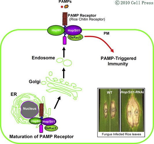 -Recognition of pathogen-associated molecular patterns represents a critical first step of innate defense in plants and animals. Ko Shimamto at NAIST and his colleagues revealed that the rice chitin receptor OsCERK1 interacts with the Hop/Sti1-Hsp90 chaperone complex in the endoplasmic reticulum(ER) and that these chaperons are required for efficient transport of OsCERK1 from the ER to the plasma membrane for chitin-triggered immunity and resistance to rice blast fungus.
| Food/Environment B6 | |
| Structure and function of environmentally-responsive proteins | |
| Ko Shimamoto | |
| News Release (in Japanese) from NAIST; | |
| Cell Host Microbe. 2010 Mar 18;7(3):185-96. The Hop/Sti1-Hsp90 chaperone complex facilitates the maturation and transport of a PAMP receptor in rice innate immunity. Chen L, Hamada S, Fujiwara M, Zhu T, Thao NP, Wong HL, Krishna P, Ueda T, Kaku H, Shibuya N, Kawasaki T, Shimamoto K. Laboratory of Plant Molecular Genetics, Nara Institute of Science and Technology, Takayama, Ikoma, Japan. |
 -Joint research team led by Keiji Tanaka at Tokyo Metropolitan Inst. Medical Science and Masayuki Yamamoto at Tohoku Univ. identifies a novel regulatory mechanism by the selective autophagy substrate p62 of the transcription factor Nrf2 through inactivation of Keap1. An verproduction of p62 or a deficiency in autophagy competes with the interaction between Nrf2 and Keap1, resulting in stabilization of Nrf2 and ranscriptional activation of Nrf2 target genes including antioxidant proteins and detoxification enzymes.
| Fundamental Biology A2: Structural basis for dynamic formation and mechanistic actions of huge and complicated proteolytic machinery(Principal Investigator: Keiji Tanaka) and Fundamental Biology B1: Structural Basis of Sensor System for Cytoprotective Gene Expression Responding to Carcinogens and Oxidative Stress (Principal Investigator: Masayuki Yamamoto) |
|
| News Release from ST - Tokyo Metropolitan Institute of Medical Science - Tohoku University; | |
| Nat Cell Biol. 2010 Feb 21. [Epub ahead of print] The selective autophagy substrate p62 activates the stress responsive transcription factor Nrf2 through inactivation of Keap1. Komatsu M, Kurokawa H, Waguri S, Taguchi K, Kobayashi A, Ichimura Y, Sou YS, Ueno I, Sakamoto A, Tong KI, Kim M, Nishito Y, Iemura S, Natsume T, Ueno T, Kominami E, Motobayashi H, Tanaka K, Yamamoto M. Laboratory of Frontier Science, Tokyo Metropolitan Institute of Medical Science, Bunkyo-ku, Tokyo 113-8613, Japan and Department of Medical Biochemistry, Tohoku University Graduate School of Medicine, 2-1 Seinyo-cho, Aoba-ku, Sendai 980-8575, Japan. PDB ID code: 3ADE |
 -Keap1 protein represses activity of the transcription factor Nrf2, and acts as a cellular sensor for oxidative stresses. Masayuki Yamamoto of Tohoku University and his colleagues reported the structure of mouse Keap1 homodimer by single particle electron microscopy. Three-dimensional reconstruction at 24-Å resolution revealed two large spheres attached by short linker arms to the sides of a small forked-stem structure, resembling a cherry-bob.
| Fundamental Biology B1 | |
| Structural Basis of Sensor System for Cytoprotective Gene Expression Responding to Carcinogens and Oxidative Stress | |
| Masayuki Yamamoto | |
| News Release from Tohoku University; | |
| Proc Natl Acad Sci USA. 2010 Jan 27. [Epub ahead of print] Keap1 is a forked-stem dimer structure with two large spheres enclosing the intervening, double glycinerepeat, and C-terminal domains. Ogura T, Tong KI, Mio K, Maruyama Y, Kurokawa H, Sato C, Yamamoto M. Department of Medical Biochemistry, Tohoku University Graduate School of Medicine, 2-1 Seinyo-cho, Aoba-ku, Sendai 980-8575, Japan. |
 -Unneeded or damaged proteins are degraded into amino acids which are used in synthesizing new proteins. Proteins are tagged for degradation with a small protein called ubiquitin. The tagging reaction is repeatedly catalyzed by enzymes called ubiquitin ligases, resulting in a polyubiquitin chain. Researchers led by Koichi Kato at Institute for Molecular Science determined crystal structure of the ubiquitin-conjugating enzyme E2 ?Eubiquitin intermediate state for ubiquitin transfer and provided structural insights into the formation of polyubiquitin chains.
| Fundamental Biology A2 | |
| Structural basis for dynamic formation and mechanistic actions of huge and complicated proteolytic machinery | |
| Keiji Tanaka | |
| News Release from Institute for Molecular Science; | |
| Structure. 2010 Jan 13;18(1):138-47. Crystal structure of UbcH5b - ubiquitin intermediate: insight into the formation of the self-assembled E2 - Ub conjugates. Sakata E, Satoh T, Yamamoto S, Yamaguchi Y, Yagi-Utsumi M, Kurimoto E, Tanaka K, Wakatsuki S, Kato K. Department of Structural Biology and Biomolecular Engineering, Graduate School of Pharmaceutical Sciences, Nagoya City University, 3-1 Tanabe-dori, Mizuho-ku, Nagoya 467-8603, Japan. PDB ID code 3A33 |
 -Researchers headed by Masaki Yamamoto at RIKEN SPring-8 Center demonstrated the successful generation of the bright micro-focused beam with 1µm width at RIKEN targeted proteins beamline (BL32XU) in SPring-8. The beamline with 1µm width could enable to determine 3-D structures of protein micro-crystals with 10µm size and is scheduled to be available for structural biologists next year.
| Structural Analysis C1 | |
| Development of the synchrotron beamlines dedicated to the measurement of micron-size protein crystals | |
| Soichi Wakatsuki | |
| News Release from RIKEN; |
 -The phytohormone abscisic acid (ABA) mediates the adaptation of plants to environmental stresses such as drought and regulates developmental signals such as seed maturation. Researchers headed by Masaru Tanokura of the University of Tokyo reported the crystal structures of the ABA receptor PYL1 bound with ABA, and the complex formed by the further binding of ABA-bound PYL1 with a group-A protein phosphatases 2C (ABI1) to reveal the ABA signalling mechanism.
| Food/Environment A3 | |
| Structural and functional analyses of transcriptional regulatory proteins useful for breading of drought and heat stress tolerant crops | |
| Masaru Tanokura | |
| News Release from the University of Tokyo; | |
| Nature. 2009 Oct 23. [Epub ahead of print] Structural basis of abscisic acid signalling. Miyazono KI, Miyakawa T, Sawano Y, Kubota K, Kang HJ, Asano A, Miyauchi Y, Takahashi M, Zhi Y, Fujita Y, Yoshida T, Kodaira K, Yamaguchi-Shinozaki K, Tanokura M. Department of Applied Biological Chemistry, Graduate School of Agricultural and Life Sciences, The University of Tokyo, Tokyo 113-8657, Japan. PDB ID code 3JRS 3JRQ |
 -Transfer RNAs are transcribed as precursor molecules which are then processed, and can undergo recoding of certain nucleotides to generate a different amino acid specificity. Osamu Nureki at the University of Tokyo and his colleagues show how the bacterial enzyme TilS uniquely recognizes and modifies a precursor tRNA Ile2 toward the mature tRNA coding for isoleucine. They demonstrate how TilS prevents the recognition of tRNAIle2 by MetRS and achieves high specificity for its substrate.
| Fundamental Biology B5 | |
| Elucidation of the mechanism of high-order cellular functions achieved by non-coding RNAs | |
| Osamu Nureki | |
| News Release from the University of Tokyo; | |
| Nature. 2009 Oct 22;461(7267):1144-8. Structural basis for translational fidelity ensured by transfer RNA lysidine synthetase. Nakanishi K, Bonnefond L, Kimura S, Suzuki T, Ishitani R, Nureki O. Department of Basic Medical Sciences, Institute of Medical Science, The University of Tokyo, 4-6-1 Shirokanedai, Minato-ku, Tokyo 108-8639, Japan PDB ID code 3A2K, 3HJ7 |
 -The gene networks that control rice flowering under long-day (LD) conditions are not well understood, even though LD flowering is agronomically important in the northern extremes of rice cultivation including Japan. Ko Shimamoto and his colleagues at Nara Institute of Science and Technology revealed that RFT1 is a florigen gene under LD conditions in rice, while Hd3a acts as a florigen gene under short-day (SD) conditions.
| Food/Environment B6 | |
| Structure and function of environmentally-responsive proteins | |
| Ko Shimamoto | |
| News Release from Nara Institute of Science and Technology; "Rice utilizes two florigen genes depending on long-day and short-day conditions" |
|
| Development. 2009 Oct;136(20):3443-50. Epub 2009 Sep 17. A gene network for long-day flowering activates RFT1 encoding a mobile flowering signal in rice. Komiya R, Yokoi S, Shimamoto K. Laboratory of Plant Molecular Genetics, Nara Institute of Science and Technology, Ikoma, Nara 630-0101, Japan |
 -Shigeyuki Yokoyama at RIKEN and his colleagues reported a dialysis-based Escherichia coli cell-free system supplemented with a combination of a steroid detergent and a natural lipid, phosphatidylcholine, for the production of a membrane protein actively integrated into liposomes. This approach might be useful for the high yield production of functional membrane proteins.
| Protein Production C1 | |
| Development of Advanced Production Technologies for Target Proteins | |
| Shigeyuki Yokoyama | |
| News Release from RIKEN; |
|
| Protein Sci. 2009 Sep 10. [Epub ahead of print] Production of functional bacteriorhodopsin by an Escherichia coli cell-free protein synthesis system supplemented with steroid detergent and lipid. Shimono K, Goto M, Kikukawa T, Miyauchi S, Shirouzu M, Kamo N, Yokoyama S. RIKEN Systems and Structural Biology Center, Kanagawa, Japan. |
 -Researchers led by Shigeyuki Yokoyama at RIKEN solved the crystal structures of aTrm5 (tRNA modification enzyme) in complex with its substrate tRNA. The structures revealed that aTrm5 has a mechanism to select L-shaped tRNAs as its substrates, and it thereby functions as a tRNA tertiary structure checkpoint in the tRNA maturation process.
| Fundamental Biology B5 | |
| Elucidation of the mechanism of high-order cellular functions achieved by non-coding RNAs | |
| Osamu Nureki | |
| News Release from RIKEN and the University of Tokyo; |
|
| Nat Struct Mol Biol. 2009 September 13. [Epub ahead of print] Tertiary structure checkpoint at anticodon loop modification in tRNA functional maturation. Goto-Ito S, Ito T, Kuratani M, Bessho Y, Yokoyama S. RIKEN Systems and Structural Biology Center, Kanagawa, Japan. PDB ID code: 2ZZM, 2ZZN |
 -Keiji Tanaka, Tokyo Metropolitan Institute of Medical Science, and his colleagues revealed that novel proteins bind specific subunits of the 19S regulatory particles (RPs) of the 26S proteasome, huge and complicated proteolytic machinery, and that these proteins are bona fide RP chaperones to help mature RPs.
| Fundamental Biology A2 | |
| Structural basis for dynamic formation and mechanistic actions of huge and complicated proteolytic machinery | |
| Keiji Tanaka | |
| News Release from Tokyo Metropolitan Organization for Medical Research; "Multiple Proteins Assist the Assembly of the 19S Regulatory Particle of the 26S Proteasome" |
|
| Cell. 2009 May 14. [Epub ahead of print] Multiple Proteasome-Interacting Proteins Assist the Assembly of the Yeast 19S Regulatory Particle. Saeki Y, Toh-E A, Kudo T, Kawamura H, Tanaka K. Tokyo Metropolitan Institute of Medical Science, Kamikitazawa, Setagaya, Japan. |
 -Keiji Tanaka, Tokyo Metropolitan Institute of Medical Science, and his colleagues identified Rfu1(Regulator for free ubiquitin chains 1) protein, which functions as an inhibitor of Doa4 (a deubiquitinating enzyme) and regulates intracellular concentrations of monomeric ubiquitins and free ubiquitin chains.
| Fundamental Biology A2 | |
| Structural basis for dynamic formation and mechanistic actions of huge and complicated proteolytic machinery | |
| Keiji Tanaka | |
| News Release from Tokyo Metropolitan Organization for Medical Research; "An Inhibitor of a Deubiquitinating Enzyme Regulates Ubiquitin Homeostasis" |
|
| Mainichi | |
| Cell. 2009 May 1;137(3):549-59. An inhibitor of a deubiquitinating enzyme regulates ubiquitin homeostasis. Kimura Y, Yashiroda H, Kudo T, Koitabashi S, Murata S, Kakizuka A, Tanaka K. Tokyo Metropolitan Institute of Medical Science, Kamikitazawa, Setagaya, Japan. |
 -Researchers headed by Yoshinori Fukui at Kyushu U. revealed, during neutrophil chemotaxis which regulates a variety of biological functions including immune response, that intracellular DOCK2 dynamics were sequentially regulated by two distinct phospholipids.
| Medicine/Pharmacology A2 | |
| Drug discovery-oriented analysis for structure and function of DOCK2 signaling molecules | |
| Yoshinori Fukui | |
| News Release from JST and Kyushu Univ.; "Deciphering the Molecular Mechanisms during Neutrophil Chemotaxis: DOCK2 Dynamics" |
|
| Nikkei | |
| Science. 2009 Mar 27. [Epub ahead of print] Sequential Regulation of DOCK2 Dynamics by Two Phospholipids during Neutrophil Chemotaxis Nishikimi A, Fukuhara H, Su W, Hongu T, Takasuga S, Mihara H, Cao Q, Sanematsu F, Kanai M, Hasegawa H, Tanaka Y, Shibasaki M, Kanaho Y, Sasaki T,. Frohman M A, and Fukui Y. Division of Immunogenetics, Department of Immunobiology and Neuroscience, Medical Institute of Bioregulation, Kyushu University, Fukuoka 812-8582, Japan |
 -Researchers led by Soichi Wakatsuki at High Energy Accelerator Research Organization(KEK) demonstrated that NEMO (NF-κB essential modulator) binding to linear ubiquitin chains plays an important role in the activation of the NF-κB pathway based on structural and functional analyses of the complex.
| Fundamental Biology A6 | |
| Structure-function analysis of protein complexes that regulate vesicular traffic | |
| Soichi Wakatsuki | |
| News Release from KEK; "Triggering an immune system - Linear Ubiquitin Chains Attach to NEMO to Activate NF-κB" |
|
| Cell. 2009 Mar 20;136(6):1098-109. Specific recognition of linear ubiquitin chains by NEMO is important for NF-kappaB activation. Rahighi S, Ikeda F, Kawasaki M, Akutsu M, Suzuki N, Kato R, Kensche T, Uejima T, Bloor S, Komander D, Randow F, Wakatsuki S, Dikic I. Structural Biology Research Center, Photon Factory, Institute of Materials Structure Science, High Energy Accelerator Research Organization (KEK), Tsukuba, Ibaraki, Japan. PDB ID code 3F89, 2ZVO, and 2ZVN |
| Medicine/Pharmacology A5 | |
| Development of anti-trypanosome drugs targeting nucleic acids and red-ox regulatory pathway | |
| Kiyoshi Kita | |
| News Release from the University of Tokyo; "Unusual and unique features in the mitochondrial respiratory enzymes from the Trypanosomatida have been identified, providing researchers with potentially invaluable targets for novel chemotherapeutic agents for Chagas disease and sleeping sickness" |
|
| J Biol Chem. 2009 Mar 13;284(11):7255-63. Epub 2009 Jan 2. Novel mitochondrial complex II isolated from Trypanosoma cruzi is composed of 12 peptides including a heterodimeric Ip subunit. Morales J, Mogi T, Mineki S, Takashima E, Mineki R, Hirawake H, Sakamoto K, Omura S, Kita K. Department of Biomedical Chemistry, Graduate School of Medicine, the University of Tokyo, Hongo, Bunkyo-ku, Tokyo, Japan. |
 -The novel technology for incorporating an iodine-labeled synthetic amino acid (3-iodo-L-tyrosine) into proteins, developed by Shigeyuki Yokoyama and his colleagues at RIKEN, could contribute to accelerate protein structure analyses.
| Protein Production C1 | |
| Development of Advanced Production Technologies for Target Proteins | |
| Shigeyuki Yokoyama | |
| News Release from RIKEN; "Iodine-labeled synthetic amino acid accelerates protein structure analyses" |
|
| Structure 2009 Mar 11;17(3):335-344 Genetic Encoding of 3-Iodo-l-Tyrosine in Escherichia coli for Single-Wavelength Anomalous Dispersion Phasing in Protein Crystallography Sakamoto K, Murayama K, Oki K, Iraha F, Kato-Murayama M, Takahashi M, Ohtake K, Kobayashi T, Kuramitsu S , Shirouzu M, Yokoyama S RIKEN Systems and Structural Biology Center, 1-7-22 Suehiro-cho, Tsurumi, Yokohama 230-0045, Japan PDB ID code 2Z0Z, 2Z10, 2Z11, 2ZXV, and 2ZP1 |
 -Researchers led by Ko Shimamoto at the Nara Institute of Science and Technology revealed that the RNA levels of Hda3 gene, encoding a floral activator (florigen), are highly correlated with flowering time, and that allelic variation of Hd1 gene is a main source of flowering time diversity in cultivated rice.
| Food/Environment B6 | |
| Structure and function of environmentally-responsive proteins | |
| Ko Shimamoto | |
| News Release from Nara Institute of Science and Technology; "Deciphering the Molecular Mechanisms Promoting the Diversity of Flowering Time in Cultivated Rice" |
|
| Asahi (asahi.com), Sankei (Sankei Digital), Nikkei | |
| Proc Natl Acad Sci U S A. 2009 Feb 25. [Epub ahead of print] Variations in Hd1 proteins, Hd3a promoters, and Ehd1 expression levels contribute to diversity of flowering time in cultivated rice. Takahashi Y, Teshima KM, Yokoi S, Innan H, and Shimamoto K. Laboratory of Plant Molecular Genetics, Nara Institute of Science and Technology, Ikoma, Nara 630-0101, Japan PMID: 19246394 |
 -Takuya Ueda and his colleagues of U. Tokyo individually synthesized the entire ensemble of Escherichia coli proteins by using an in vitro reconstituted translation system and analyzed the aggregation propensities. A histogram of the solubilities revealed a clear bimodal distribution.
| Protein Production C1 | |
| Development of Advanced Production Technologies for Target Proteins | |
| Shigeyuki Yokoyama | |
| News Release from Univ. Tokyo; "Bimodal protein solubility distribution of the entire ensemble of Escherichia coli proteins" |
|
| Biotechnology Japan Online (February 27) | |
| Proc Natl Acad Sci U S A. 2009 Feb 27. [Epub ahead of print] Bimodal protein solubility distribution revealed by an aggregation analysis of the entire ensemble of Escherichia coli proteins. Niwa T, Ying BW, Saito K, Jin W, Takada S, Ueda T, Taguchi H. Department of Medical Genome Sciences, Graduate School of Frontier Sciences, University of Tokyo, Kashiwa, Chiba 277-8562, Japan PMID: 19251648 |
 -Researchers headed by Junn Yanagisawa at the University of Tsukuba have discovered that an enzyme called CHIP, which is known to degrade a number of cancer causing proteins, acts to degrade the gene regulator SRC-3, which then suppresses tumor progression in breast cancer.
| Medicine/Pharmacology B2Food/Environment A4 | |
| Development of new compounds for fibrosis therapy | |
| Junn Yanagisawa | |
| News Release from Tsukuba Univ.; "CHIP enzyme prevents breast tumor growth and metastasis" |
|
| Yomiuri, Mainichi, Reuters, ecancermedicalscience | |
| Nat Cell Biol. 2009 Feb 8. [Epub ahead of print] The ubiquitin ligase CHIP acts as an upstream regulator of oncogenic pathways. Kajiro M, Hirota R, Nakajima Y, Kawanowa K, So-Ma K, Ito I, Yamaguchi Y, Ohie SH, Kobayashi Y, Seino Y, Kawano M, Kawabe YI, Takei H, Hayashi SI, Kurosumi M, Murayama A, Kimura K, Yanagisawa J. Graduate School of Life and Environmental Sciences, University of Tsukuba, Tsukuba Science City, Ibaraki 305-8572, Japan. PMID: 19198599 |
| Fundamental Biology B5 | |
| Elucidation of the mechanism of high-order cellular functions achieved by non-coding RNAs | |
| Osamu Nureki | |
| News Release from JST and U. Tokyo; "Pyrrolysyl-tRNA synthetase-tRNA structure reveals the molecular basis of orthogonality" |
|
| Nature. 2009 Feb 26;457(7233):1163-7. Epub 2008 Dec 31. Pyrrolysyl-tRNA synthetase-tRNA(Pyl) structure reveals the molecular basis of orthogonality. Nozawa K, O'Donoghue P, Gundllapalli S, Araiso Y, Ishitani R, Umehara T, Soll D, Nureki O. Department of Basic Medical Sciences, Institute of Medical Science, The University of Tokyo, 4-6-1 Shirokanedai, Minato-ku, Tokyo 108-8639, Japan PMID: 19118381 PDB ID code: 2ZNJ, 2ZNI |
 -The structure of receptor GID1 of key plant hormone gibberellin, reported by Makoto Matsuoka, a plant scientist at Nagoya Univ., and Hiroaki Kato, a structural biologist at Kyoto Univ., reveals how the hormone works.
| Food/Environment A4 | |
| Structural and functional analyses of regulatory proteins in plant growth and stress resistance, that is useful for improvement of the stress-resistant | |
| Makoto Matsuoka | |
| News Release from Nagoya Univ., Kyoto Univ. and RIKEN; "Structure of gibberellin revealed - it could help in designing more potent plant growth regulators for the second green revolution era" |
|
| Asahi, Yomiuri, Nikkan-Kogyo (November 27), Chemical & Engineering News (December 1) | |
| Nature. 2008 Nov 27;456 (7221)::520-523. Structural basis for gibberellin recognition by its receptor GID1 Shimada A, Ueguchi-Tanaka M, Nakatsu T, Nakajima M, Naoe Y, Ohmiya H, Kato H, Matsuoka M. Bioscience and Biotechnology Center, Nagoya University, Nagoya, Aichi 464-8601, JapanPMID: 19037316 [PubMed - in process] PDB ID code 3EBL and 3ED1 |
| Fundamental Biology B5 | |
| Elucidation of the mechanism of high-order cellular functions achieved by non-coding RNAs | |
| Osamu Nureki | |
| News Release from U. Tokyo; "Structural transition of protein secretion machinery across membranes" |
|
| Nature. 2008 Oct 16;455(7215):988-91. Conformational transition of Sec machinery inferred from bacterial SecYE structures. Tsukazaki T, Mori H, Fukai S, Ishitani R, Mori T, Dohmae N, Perederina A, Sugita Y, Vassylyev DG, Ito K, Nureki O. Department of Basic Medical Sciences, Institute of Medical Science, The University of Tokyo, 4-6-1 Shirokanedai, Minato-ku, Tokyo 108-8639, Japan PMID: 18923527 PDB ID code: 2ZJS, 2ZQP |
| Protein Production C1 | |
| Development of Advanced Production Technologies for Target Proteins. | |
| Shigeyuki Yokoyama | |
| News Release from RIKEN and Univ. Tokyo; "Novel fused enzyme which selectively incorporates nonnatural amino acid into proteins - an editing-domain transplanted tyrosyl-tRNA synthetase with enhanced specificity" |
|
| Nikkei, Nikkei-Sangyo (August 20) | |
| Proc Natl Acad Sci U S A. 2008 Sep 9;105(36):13298-303. Epub 2008 Sep 2. Transplantation of a tyrosine editing domain into a tyrosyl-tRNA synthetase variant enhances its specificity for a tyrosine analog. Oki K, Sakamoto K, Kobayashi T, Sasaki HM, Yokoyama S. Department of Biophysics and Biochemistry, Graduate School of Science, University of Tokyo, 7-3-1 Hongo, Bunkyo-ku, Tokyo 113-0033, Japan. PMID: 18765802 [PubMed - - indexed for MEDLINE] |
 -Researchers at Yokohama City University headed by Yoshio Goshima and Zenro Ikezawa found that semaphorin3a alleviates skin lesions and scratching behavior in mice.
| Medicine / Pharmacology B4 | |
| Structural and functional analysis of semaphorins and their receptors. | |
| Atsushi Kumanogoh | |
| News Release from Yokohama City University; "Semaphorin3A, a potent inhibitor of neurite outgrowth, is promising in the treatment of atopic dermatitis" |
|
| Mainichi (July 22) | |
| J Invest Dermatol. 2008 Dec;128(12):2842-9. Epub 2008 Jul 10 Semaphorin3A Alleviates Skin Lesions and Scratching Behavior in NC/Nga Mice, an Atopic Dermatitis Model. Yamaguchi J, Nakamura F, Aihara M, Yamashita N, Usui H, Hida T, Takei K, Nagashima Y, Ikezawa Z, Goshima Y. Department of Molecular Pharmacology and Neurobiology, Yokohama City University Graduate School of Medicine, Yokohama, Japan. PMID: 18615113 [PubMed - in process] |
| Fundamental Biology B4 | |
| Towards structure-based design of novel inhibitors for V -ATPase | |
| So Iwata | |
| News Release from Japan Science and Technology Agency, RIKEN and Kyoto Univ.; "Transport mechanism at the rotor ring of sodium ion transporting enzyme V-ATPase was elucidated" |
|
| - | |
| Proc Natl Acad Sci U S A. 2008 Jun 24;105(25):8607-12. Epub 2008 Jun 16. Ion binding and selectivity of the rotor ring of the Na+-transporting V-ATPase. Murata T, Yamato I, Kakinuma Y, Shirouzu M, Walker JE, Yokoyama S, Iwata S. Department of Cell Biology, Faculty of Medicine, Kyoto University, Yoshidakonoe-cho, Sakyo-ku, Kyoto 606-8501, Japan. PMID: 18559856 [PubMed - indexed for MEDLINE] PDB ID code 2CYD |
 -The specific labeling of proteins in living cells using a genetically encodable tag and a small synthetic probe targeting the tag has been created. This new probe reported by Katsumi Matsuzaki, et al. of Kyoto University was featured in several Japanese newspapers.
| Chemical Regulation C1 | |
| Establishment of Chemical Library and Development of Protein Regulation Technology | |
| Tetsuo Nagano | |
| News Release from Kyoto Univ.; "Quick labeling of drug receptors with diverse fluorophores - highly tractable and promising method for visualization of membrane receptors" |
|
| Kyoto, Sankei (June 14), Nikkan-Kogyo, Nikkei-Sangyo (June 16) | |
| ACS Chem Biol. 2008 Jun 20;3(6):341-5. Coiled-coil tag--probe system for quick labeling of membrane receptors in living cell. Yano Y, Yano A, Oishi S, Sugimoto Y, Tsujimoto G, Fujii N, Matsuzaki K. Graduate School of Pharmaceutical Sciences, Kyoto University, Kyoto, Japan. PMID: 18533657 |